. 2012;7(7):e40463.
doi: 10.1371/journal.pone.0040463. Epub 2012 Jul 12.

STAT1 interacts with RXRα to upregulate ApoCII gene expression in macrophages

Affiliations

STAT1 interacts with RXRα to upregulate ApoCII gene expression in macrophages

Violeta G Trusca et al. PLoS One. 2012.

Abstract

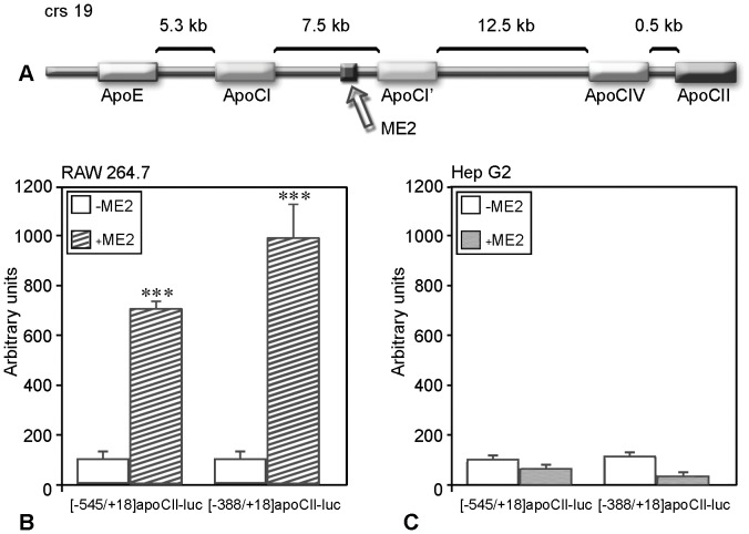
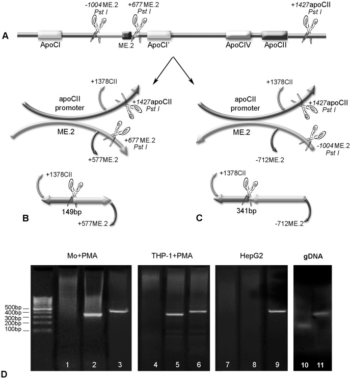
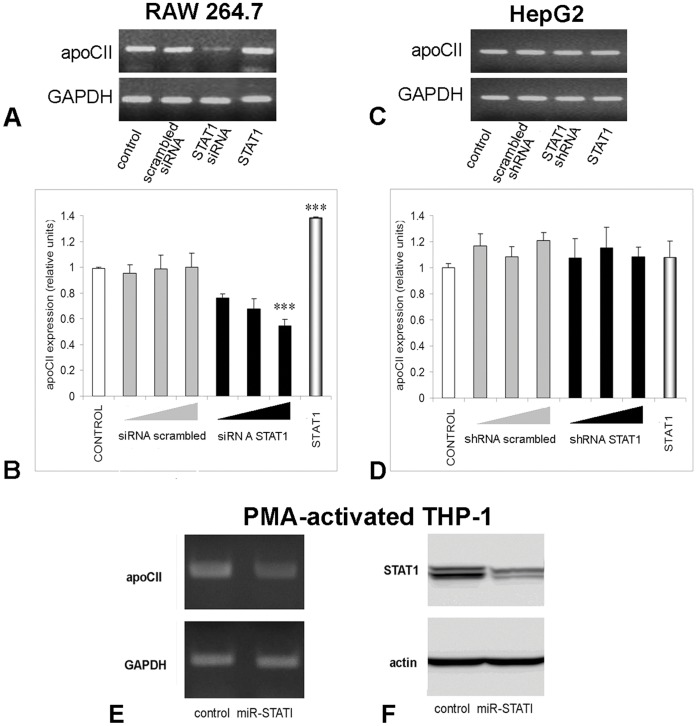
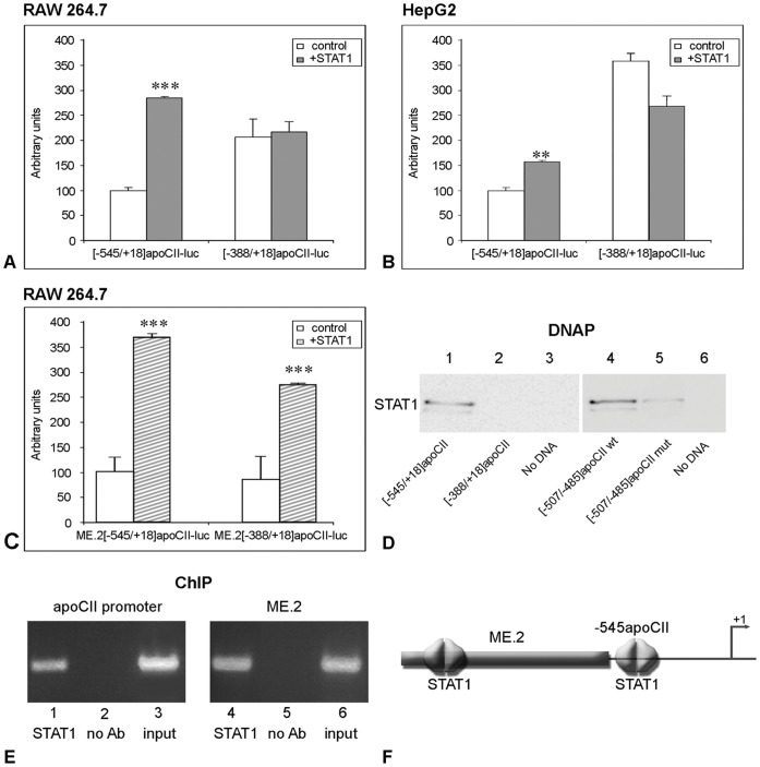
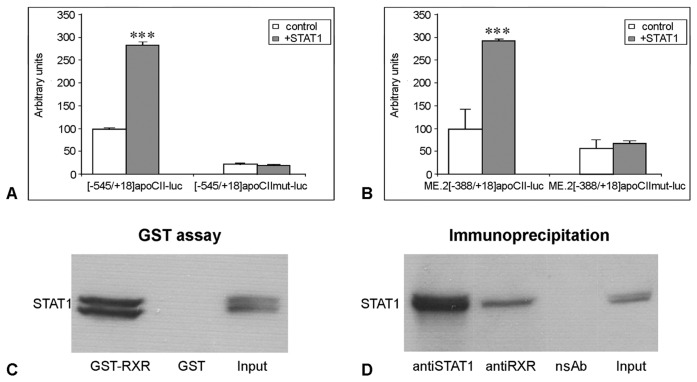
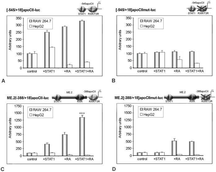
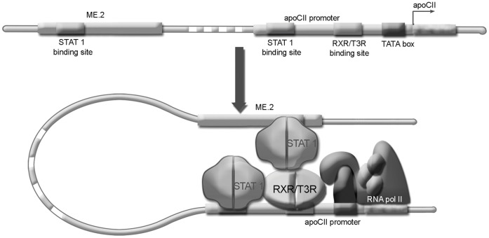
Apolipoprotein CII (apoCII) is a specific activator of lipoprotein lipase and plays an important role in triglyceride metabolism. The aim of our work was to elucidate the regulatory mechanisms involved in apoCII gene modulation in macrophages. Using Chromosome Conformation Capture we demonstrated that multienhancer 2 (ME.2) physically interacts with the apoCII promoter and this interaction facilitates the transcriptional enhancement of the apoCII promoter by the transcription factors bound on ME.2. We revealed that the transcription factor STAT1, previously shown to bind to its specific site on ME.2, is functional for apoCII gene upregulation. We found that siRNA-mediated inhibition of STAT1 gene expression significantly decreased the apoCII levels, while STAT1 overexpression in RAW 264.7 macrophages increased apoCII gene expression. Using transient transfections, DNA pull down and chromatin immunoprecipitation assays, we revealed a novel STAT1 binding site in the -500/-493 region of the apoCII promoter, which mediates apoCII promoter upregulation by STAT1. Interestingly, STAT1 could not exert its upregulatory effect when the RXRα/T3Rβ binding site located on the apoCII promoter was mutated, suggesting physical and functional interactions between these factors. Using GST pull-down and co-immunoprecipitation assays, we demonstrated that STAT1 physically interacts with RXRα. Taken together, these data revealed that STAT1 bound on ME.2 cooperates with RXRα located on apoCII promoter and upregulates apoCII expression only in macrophages, due to the specificity of the long-range interactions between the proximal and distal regulatory elements. Moreover, we showed for the first time that STAT1 and RXRα physically interact to exert their regulatory function.


Conflict of interest statement

Competing Interests: The authors have declared that no competing interests exist.

Figures

Figure 1
Figure 1. ApoCII promoter activity is upregulated by ME.2 in macrophages but not in hepatocytes.
Panel A. Schematic representation of the apoE/apoCI/apoCI’/apoCIV/apoCII gene cluster and the location of ME.2. Panels B and C. RAW 264.7 macrophages and HepG2 cells were transiently transfected with plasmids in which [−545/+18]apoCII or [−388/+18]apoCII promoter fragments were cloned in the presence (+ME.2) or in the absence (–ME.2) of ME.2 in pGL3 basic vector. In RAW 264.7 macrophages, both promoter fragments activities can be enhanced by ME.2 (Panel B) while in HepG2 hepatocytes, ME.2 cannot increase apoCII promoter activity (Panel C).
Figure 2
Figure 2. Interactions between apoCII promoter and ME.2 in macrophages as revealed by chromosome conformation capture (3C) assays.
Panels A–C. Schematic illustration of the linear alignment of ME.2 and apoCII (including the Pst I sites adjacent to these regions) and the possible interactions between apoCII promoter and ME.2 in sense (B) or antisense (C) orientation. For 3C experiments, the PstI restriction enzyme was used. PstI restriction sites are located at positions +1427 in apoCII gene as well as at +677 bp downstream of ME.2 and at −1004 bp upstream of ME.2, as showed in the picture. The primers flanking the newly ligated fragments are also illustrated: for both cases (B and C), the forward primer was +1378CII, and the reverse primers were: +577ME.2 (B) and −712ME.2 (C). The predicted lengths of the ligated fragments are: 149 bp for “sense orientation” (B) and 341 bp and “antisense orientation” interaction with ME.2 (C). Panel D. Human peripheral blood monocytes (lanes 1–3) and THP-1 cells (lanes 4–6) differentiated to macrophages by PMA treatment, as well as HepG2 cells (lanes 7–9), were subjected to the 3C assay. PCR fragments amplified with primers +1378CII and +577ME.2 (testing the interaction in sense orientation) are shown in lanes 1, 4 and 7, and PCR products obtained with primers +1378CII and −712ME.2 (testing the interaction in antisense orientation) are shown in lanes 2, 5 and, respectively 8. The positive control for DNA integrity and PCR (amplification of a 468 bp fragment of ME.2) are shown in lanes 3, 6 and respectively 9. ME.2 and the apoCII promoter interact in antisense orientation since only the fragment of 341 bp was obtained from human monocytes and THP-1 cells differentiated with PMA (lane 2 and, respectively 5) whereas no bands were obtained for sense orientation interaction (lanes 1 and, respectively 4). In contrast, in HepG2 cells, 3C experiments gave no bands either for sense or antisense orientation interaction (lanes 7 and, respectively 8), revealing that apoCII promoter and ME.2 do not interact in hepatocytes. As control, genomic DNA isolated from THP-1 cells was used, and the amplification of the ligation products using +1378CII and +577ME.2 primers, and +1378CII and −712ME.2 primers, gave the expected size of the PCR products of 149 bp and 341 bp (Fig. 2D, lanes 10 and 11, respectively).
Figure 3
Figure 3. STAT1 positively modulates apoCII expression in macrophages, but not in hepatocytes.
To determine apoCII modulation by STAT1 silencing, RAW 264.7 cells were transfected with siRNA specific for mouse STAT1 (Panel A and B), in HepG2 cells were transfected with STAT1 shRNA producing vector (Panel C and D), and in PMA-activated THP-1 cells were transduced with a lentiviral expression vector for a micro RNA cassette for human STAT1 (Panel E). ApoCII modulation was tested by RT-PCR (A, C and E) and quantified by Real Time PCR (B and D). GAPDH expression was tested to normalize the apoCII expression. The results showed that apoCII expression was strongly inhibited in macrophages (Panel A, lane STAT1 siRNA and Panel E lane miR-STAT1), but not in hepatocytes (Panel C, lane STAT1 shRNA). Real Time PCR experiments showed a dose-dependent inhibition of apoCII by STAT1 siRNA in RAW 264.7 macrophages (for the highest dose, p<0.005), but not in HepG2 cells (Panels B, and D, respectively, black columns). As control, we used scrambled siRNA for RAW.264.7 cells, or scrambled shRNA for HepG2 cells, which did not affect significantly the apoCII expression. STAT1 expression was inhibited more than 50% by transduction of the PMA-activated THP1 with miR-STAT1, as detected by Western blotting (Panel F). To determine apoCII modulation by STAT1 overexpression, RAW 264.7 macrophages and HepG2 cells were transfected with a STAT1 expression vector and subjected to RT-PCR. ApoCII mRNA level was increased in STAT1-overexpressing macrophages (Panel A, lane STAT1), but not in hepatocytes (Panel C lane STAT1, and Panel D last column). Real Time PCR experiments revealed an increase of ∼1.4 folds in apoCII expression in STAT1 overexpressing macrophages (Panels B, last columns; p<0.005).
Figure 4
Figure 4. STAT1-dependent transactivation of apoCII promoter in RAW 264.7 cells.
Panel A, B. RAW 264.7 macrophages (A) and HepG2 hepatocytes (B) were transiently transfected with plasmids containing [−545/+18] or [−388/+18]apoCII promoter fragments in the presence or in the absence (control) of STAT1 expression vector. STAT1 overexpression increased the activity of [−545/+18]apoCII promoter in RAW 264.7 cells (∼3.4 fold, p<0.0001) as well as in HepG2 cells (∼1.5 fold, p<0.002). By contrast, the activity of the [−388/+18]apoCII promoter was not increased by STAT1 overexpression, either in macrophages (p>0.5) or in hepatocytes. Panel C. RAW 264.7 macrophages were transfected with ME.2[−545/+18]apoCII-luc or with ME.2[−388/+18]apoCII-luc plasmids in the absence (control) or in the presence of STAT1 expression vector. In the presence of ME.2, the activities of [−545/+18] and [−388/+18] promoter fragments were increased by STAT1 overexpression (3.8 fold, p<0.0002 and 2.9 fold, respectively p<0.0002). Panel D. DNA pull down assays were performed using different fragments ([−545/+18] and [−388/+18]) of apoCII promoter or oligonucleotides containing the wild type or mutated [−507/−485] region of apoCII promoter, and nuclear extracts obtained from STAT1- overexpressing HepG2 (right) and RAW 264.7 (left) cells. The results showed that the [−545/+18]apoCII promoter, but not [−388/+18] fragment can bind STAT1 proteins (lane 1 and, respectively 2). STAT1 is able to bind the wild type [−507/−485] region of apoCII promoter (lane 4), while STAT1 binding to the mutated region is impaired (lane 5). Negative controls, in which DNAP was performed without DNA, are illustrated in lanes 3 and 6 (no DNA). Panel E. ChIP experiments performed using anti-STAT1 antibodies showed that STAT1 is recruited to the [−738/−336]apoCII gene fragment as well as to the [140–440] ME.2 region (lanes 1 and, respectively, 4). No bands were obtained when anti-STAT1 antibodies were omitted in ChIP experiments for apoCII promoter and for ME.2 region (lanes 2 and, respectively, 5). PCR using the input as template and primers for apoCII promoter or ME.2 gave the expected bands (lanes 3 and, respectively, 6). Panel F. Schematic representation of STAT1 binding sites on apoCII proximal promoter and ME.2 region.
Figure 5
Figure 5. STAT1 interacts with RXRα to modulate apoCII promoter activity.
Panel A, B. RAW 264.7 macrophages were transiently transfected with [−545/+18]apoCII-luc (A) and ME.2[−388/+18]apoCII-luc (B) or with their corresponding mutants [−545/+18]apoCIImut-luc (A) and ME.2[388/+18]apoCIImut-luc (B) in the presence or in the absence (control) of STAT1 expression vector. The mutation in RXRα/T3Rβ site located at −152/−135 in apoCII promoter impaired the upregulatory effect of STAT1 that could bind to the −500/−493 region of the apoCII promoter (A) or to the 174–182 region of ME.2 (B). Panel C. GST-pull down assay performed using cellular extracts from STAT1-overexpressing RAW 264.7 macrophages indicated that STAT1 proteins bind efficiently to the GST-RXRα-beads (lane GST-RXRα), but cannot bind to the GST beads (lane GST); Western blotting for STAT1 using the whole cell extract is illustrated in the lane ‘Input’. Panel D. Co-immunoprecipitation experiments were performed using cellular extracts from HEK-293T cells overexpressing both STAT1 and RXRα. The antibodies used for immunoprecipitation are indicated (anti-STAT1 and anti-RXRα). Western blotting of immunoprecipitated proteins done with anti-STAT1 antibodies revealed that STAT1 proteins were efficiently immunoprecipitated by anti-STAT1 antibodies (lane anti-STAT1), but they were also co-immunoprecipitated using anti-RXRα antibodies (lane anti-RXRα). No bands were obtained in the negative control, in which the samples were subjected to immunoprecipitation in the absence of the antibodies (lane NoAb). Western blot for STAT1 using whole cell extract is presented in the lane input.
Figure 6
Figure 6. Transactivation of apoCII promoter by STAT1 is amplified by RXRα ligand in macrophages, but not in hepatocytes.
Panel A – D. RAW 264.7 macrophages and HepG2 hepatocytes were transiently transfected with the constructs [−545/+18]apoCII-luc, [−545/+18]apoCIImut-luc, ME.2[−388/+18]apoCII-luc, and ME.2[−388/+18]apoCIImut-luc under basal condition (control), in the presence of STAT1 overexpression (STAT1), after 9-cis-retinoic acid treatment (RA) or in the presence of both activators (STAT1+RA). The concomitant STAT1 overexpression and RA treatment of RAW 264.7 macrophages enhanced the wild type apoCII promoter at a higher level than each activator alone (Panel A, grey columns). This effect on apoCII promoter was more pronounced when the promoter was under the control of ME.2 (Panel C, grey columns). The activity of the mutant apoCII promoter was not affected by either activator alone (STAT1 or RA) or in combination (Panel B, grey columns). In the presence of ME.2, RA treatment of RAW 264.7 cells increased the activity of the mutant apoCII promoter (due to the RXR binding sites found on ME.2), but STAT1 could not potentiate this enhancement (Panel D, grey columns). By contrast, in HepG2 cells the cumulative effect of STAT1 and RXR was not observed on any constructs employed (white columns).
Figure 7
Figure 7. Schematic representation showing STAT1/RXRα interactions in apoCII gene regulation.
In macrophages, the ME.2 is brought in the proximity of apoCII promoter. This DNA bending facilitates the interaction of STAT1 attached on its site found on ME.2 with RXRα bound on the apoCII promoter leading to apoCII gene transactivation. A similar mechanism of apoCII transactivation involving interaction of STAT1 with RXRα takes place when STAT1 is attached on its site found on apoCII promoter. After STAT1-RXRα interaction, this complex may cooperate with the basal transcription factors of the initiation transcription complex, thus leading to apoCII gene activation.

Similar articles

Cited by

References

    1. Jackson RC, Blobel G. Post-translational cleavage of presecretory proteins with an extract of rough microsomes from dog pancreas containing signal peptidase activity. Proc Natl Acad Sci U S A. 1977;74:5598–5602. - PMC - PubMed
    1. Miller AL, Smith LC. Activation of lipoprotein lipase by apolipoprotein glutamic acid. Formation of a stable surface film. J Biol Chem. 1973;248:3359–3362. - PubMed
    1. Zannis VI, Kardassis D, Zanni EE. Genetic mutations affecting human lipoproteins, their receptors, and their enzymes. Adv Hum Genet. 1993;21:145–319. - PubMed
    1. Nilsson-Ehle P, Garfinkel AS, Schotz MC. Lipolytic enzymes and plasma lipoprotein metabolism. Annu Rev Biochem. 1980;49:667–693. - PubMed
    1. Myklebost O, Williamson B, Markham AF, Myklebost SR, Rogers J, et al. The isolation and characterization of cDNA clones for human apolipoprotein CII. J Biol Chem. 1984;259:4401–4414. - PubMed

Publication types