. 2012;7(7):e40761.
doi: 10.1371/journal.pone.0040761. Epub 2012 Jul 10.

Campylobacter jejuni induces acute enterocolitis in gnotobiotic IL-10-/- mice via Toll-like-receptor-2 and -4 signaling

Affiliations

Campylobacter jejuni induces acute enterocolitis in gnotobiotic IL-10-/- mice via Toll-like-receptor-2 and -4 signaling

Lea-Maxie Haag et al. PLoS One. 2012.

Abstract

Background: Campylobacter jejuni is a leading cause of foodborne bacterial enterocolitis worldwide. Investigation of immunopathology is hampered by a lack of suitable vertebrate models. We have recently shown that gnotobiotic mice as well as conventional IL-10(-/-) animals are susceptible to C. jejuni infection and develop intestinal immune responses. However, clinical symptoms of C. jejuni infection were rather subtle and did not reflect acute bloody diarrhea seen in human campylobacteriosis.

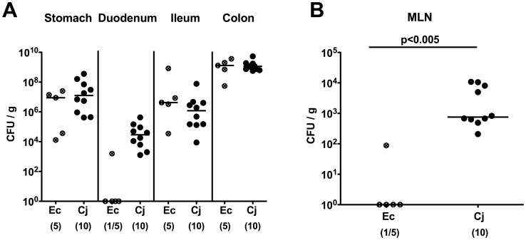
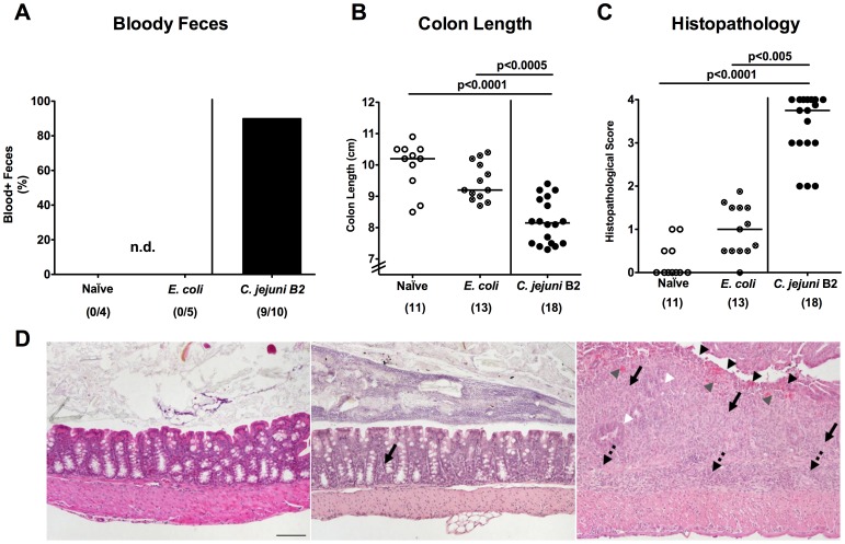
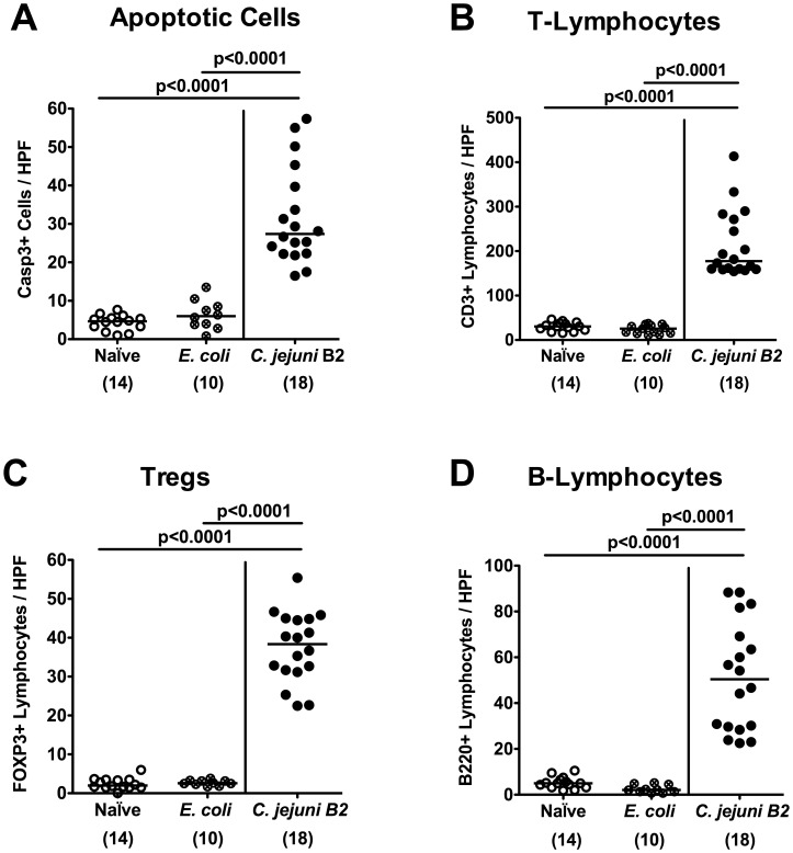
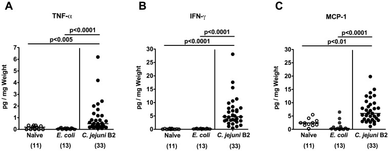
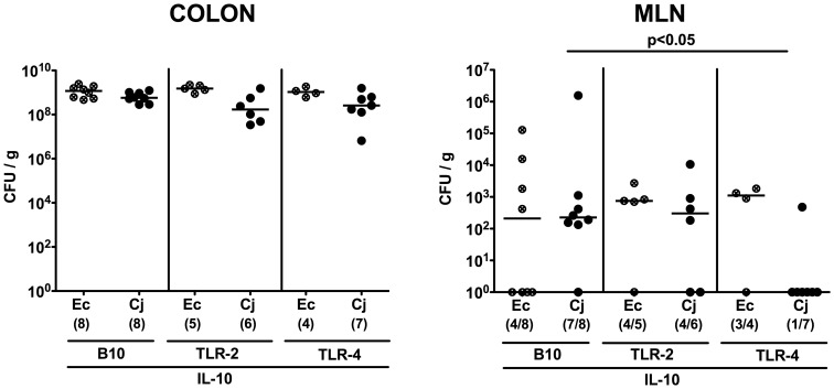
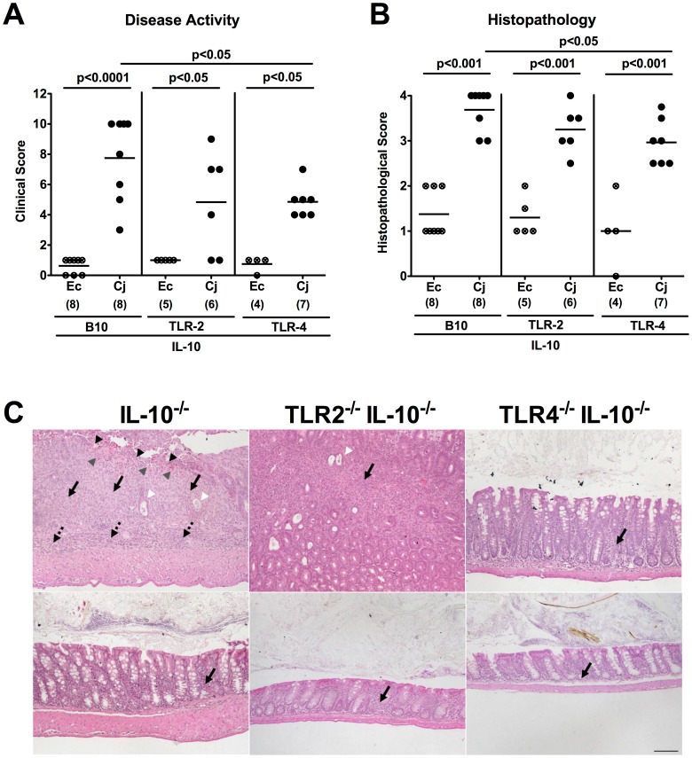
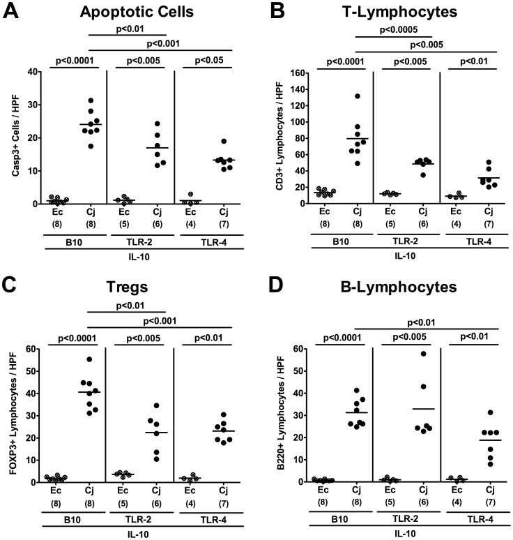
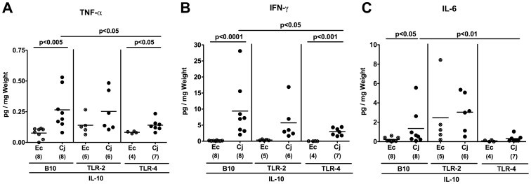
Methodology/principal findings: In order to overcome these limitations we generated gnotobiotic IL-10(-/-) mice by quintuple antibiotic treatment starting right after weaning. The early treatment was essential to prevent these animals from chronic colitis. Following oral infection C. jejuni colonized the gastrointestinal tract at high levels and induced acute enterocolitis within 7 days as indicated by bloody diarrhea and pronounced histopathological changes of the colonic mucosa. Immunopathology was further characterized by increased numbers of apoptotic cells, regulatory T-cells, T- and B-lymphocytes as well as elevated TNF-α, IFN-γ, and MCP-1 concentrations in the inflamed colon. The induction of enterocolitis was specific for C. jejuni given that control animals infected with a commensal E. coli strain did not display any signs of disease. Most strikingly, intestinal immunopathology was ameliorated in mice lacking Toll-like-receptors-2 or -4 indicating that C. jejuni lipoproteins and lipooligosaccharide are essential for induction and progression of immunopathology.

Conclusion/significance: Gnotobiotic IL-10(-/-) mice develop acute enterocolitis following C. jejuni infection mimicking severe episodes of human campylobacteriosis and are thus well suited to further dissect mechanisms underlying Campylobacter infections in vivo.


Conflict of interest statement

Competing Interests: Co-authors Stefan Bereswill and Markus M. Heimesaat are PLoS ONE Editorial Board members. This does not alter the authors' adherence to all the PLoS ONE policies on sharing data and materials.

Figures

Figure 1
Figure 1. C. jejuni B2 colonization and translocation in gnotobiotic IL-10−/− mice.
Gnotobiotic IL-10−/− mice were generated by antibiotic treatment and orally infected either with C. jejuni B2 (solid circles) or an apathogenic E. coli strain (crossed circles) derived from the commensal gut microbiota of conventional colonized wildtype mice as described in Methods. C. jejuni B2 and E. coli loads were determined in luminal samples taken from different compartments of the gastrointestinal tract (A) and mesenteric lymphnodes (MLN) (B) at day 7 p.i. (CFU, colony forming units). Numbers of animals harboring the respective bacterial species out of the total number of analyzed animals are given in parentheses. Medians (black bars) and significance levels (P-values) determined by Mann-Whitney-U test are indicated. Data shown are representative for three independent experiments.
Figure 2
Figure 2. C. jejuni induces acute enterocolitis in gnotobiotic IL-10−/− mice.
Gnotobiotic IL-10−/− mice were generated by antibiotic treatment and orally infected either with C. jejuni B2 (solid circles; black bar) or a commensal E. coli strain (crossed circles) derived from the commensal gut microbiota of conventional colonized wildtype mice as described in Methods. Uninfected gnotobiotic IL-10−/− mice served as negative controls (open circles). (A) Relative rate of blood-positive stool samples of infected IL-10−/− and uninfected control mice were determined by haemoccult test at day 7 p.i. (see Methods). Numbers of animals with haemcoocult positive stool samples out of the total number of analyzed animals are given in parentheses (n.d., not detectable). (B) Absolute colon lengths and (C) histopathological changes applying a standardized histopathological score in HE-stained colonic paraffin sections were determined at day 7 p.i., illustrated by (D) representative photomicrographs out of three independent experiments (100x magnification, scale bar 100 µm) derived from naïve (left), E. coli- (middle) and C. jejuni B2- (right) infected animals. Solid arrows indicate mucosal, dotted arrows submucosal infiltrates, black arrow heads ulcerations, white arrow heads loss of goblet cells, and gray arrow heads mucosal bleeding. Numbers of analyzed animals are given in parentheses. Medians (black bars) and significance levels (P-values) determined by Mann-Whitney-U test are indicated. Data shown were pooled from at least three independent experiments.
Figure 3
Figure 3. Inflammatory and immune cell responses following C. jejuni infection of gnotobiotic IL-10−/− mice.
Gnotobiotic IL-10−/− mice were generated by antibiotic treatment and orally infected either with C. jejuni B2 (solid circles) or an apathogenic E. coli strain (crossed circles) derived from the commensal gut microbiota of conventional colonized wildtype mice as described in Methods. Uninfected gnotobiotic IL-10−/− mice served as negative controls (open circles). The average numbers of apoptotic cells (positive for caspase-3, panel A), T-lymphocytes (positive for CD3, panel B), regulatory T-cells (Tregs, positive for FOXP3, panel C) and B-lymphocytes (positive for B220, panel D) from at least six high power fields (HPF, 400x magnification) per animal were determined microscopically in immunohistochemically stained colon sections at day 7 p.i. Numbers of analyzed animals are given in parentheses. Means (black bars) and levels of significance (P-values) determined by the Student’s t-test are indicated. Data shown are pooled from at least three independent experiments.
Figure 4
Figure 4. Pro-inflammatory cytokine responses in the colon of C. jejuni infected gnotiotic IL-10−/− mice.
Gnotobiotic IL-10−/− mice were generated by antibiotic treatment and orally infected with either C. jejuni B2 (solid circles) or an apathogenic E. coli strain (crossed circles) derived from the commensal gut microbiota of conventional colonized wildtype mice as described in Methods. Uninfected gnotobiotic IL-10−/− mice served as negative controls (open circles). Concentrations (pg per mg colon) of (A) TNF-α, (B) IFN-γ, and (C) MCP-1 were determined in supernatants of ex vivo colonic cultures at day 7 p.i. by cytometric bead array (CBA). Numbers of analyzed animals are given in parentheses. Means (black bars) and levels of significance (P-values) as compared to the respective group (determined by Student’s t-test) are indicated. Data shown are pooled from at least three independent experiments.
Figure 5
Figure 5. TLR-dependent C. jejuni colonization and translocation in gnotobiotic IL-10−/− mice.
Gnotobiotic IL-10−/− (B10 background) as well as TLR-2−/− IL-10−/− and TLR-4−/− IL-10−/− mice were generated by antibiotic treatment and orally infected either with C. jejuni B2 (Cj; solid symbols) or a commensal E. coli strain (Ec; crossed symbols) as described in Methods. C. jejuni B2 and E. coli loads were determined in luminal colonic samples (A) and mensenteric lymphnodes (MLN) (B) at day 7 p.i. (CFU, colony forming units). Numbers of animals harboring the respective bacterial species out of the total number of analyzed animals are given in parentheses. Medians (black bars) and significance levels (P-values) determined by Mann-Whitney-U test are indicated. Data shown are representative for three independent experiments.
Figure 6
Figure 6. C. jejuni induces TLR-dependent acute enterocolitis in gnotobiotic IL-10−/− mice.
Gnotobiotic IL-10−/− (B10 background) as well as TLR-2−/− IL-10−/− and TLR-4−/− IL-10−/− mice were generated by antibiotic treatment and orally infected either with C. jejuni B2 (Cj; solid symbols) or a commensal E. coli strain (Ec; crossed symbols) derived from the commensal gut microbiota of conventional colonized wildtype mice as described in Methods. (A) Disease activity of infected mice and (B) histopathological changes in HE-stained colonic paraffin sections applying a standardized clinical and histopathological score, respectively, were determined at day 7 p.i., illustrated by (C) representative photomicrographs out of three independent experiments (100x magnification, scale bar 100 µm) derived from C. jejuni B2- (upper panel) and E. coli- (lower panel) infected IL-10−/− (left panel), TLR-2−/− IL-10−/− (middle panel), and TLR-4−/− IL-10−/− (right panel) mice. Solid arrows indicate mucosal, dotted arrows submucosal iinfiltrates, black arrow heads ulcerations, white arrow heads crypt drop-out, and gray arrow heads mucosal bleeding. Numbers of analyzed animals are given in parentheses. Medians (black bars) and significance levels (P-values) determined by Mann-Whitney-U test are indicated. Data shown are representative for three independent experiments.
Figure 7
Figure 7. TLR-dependent inflammatory and immune cell responses following C. jejuni infection of gnotobiotic IL-10−/− mice.
Gnotobiotic IL-10−/− (B10 background) as well as TLR-2−/− IL-10−/− and TLR-4−/− IL-10−/− mice were generated by antibiotic treatment and orally infected either with C. jejuni B2 (Cj; solid symbols) or a commensal E. coli strain (Ec; crossed symbols) as described in Methods. The average numbers of apoptotic cells (positive for caspase-3, panel A), T-lymphocytes (positive for CD3, panel B), regulatory T-cells (Tregs, positive for FOXP3, panel C) and B-lymphocytes (positive for B220, panel D) from at least six high power fields (HPF, 400x magnification) per animal were determined microscopically in immunohistochemically stained colon sections at day 7 p.i. Numbers of analyzed animals are given in parentheses. Means (black bars) and levels of significance (P-values) determined by the Student’s t-test are indicated. Data shown are representative for three independent experiments.
Figure 8
Figure 8. TLR-dependent pro-inflammatory cytokine responses in the colon of C. jejuni infected gnotobiotic IL-10−/− mice.
Gnotobiotic IL-10−/− (B10 background) as well as TLR-2−/− IL-10−/− and TLR-4−/− IL-10−/− mice were generated by antibiotic treatment and orally infected either with C. jejuni B2 (Cj; solid symbols) or a commensal E. coli strain (Ec; crossed symbols) as described in Methods. Concentrations (pg per mg colon) of (A) TNF-α, (B) IFN-γ, and (C) IL-6 were determined in supernatants of ex vivo colon cultures at day 7 p.i. by cytometric bead array (CBA). Numbers of analyzed animals are given in parentheses. Means (black bars) and levels of significance (P-values) as compared to the respective group (determined by Student’s t-test) are indicated. Data shown are representative for three independent experiments.

References

    1. Hermans D, Pasmans F, Messens W, Martel A, Van Immerseel F, et al. Poultry as a Host for the Zoonotic Pathogen Campylobacter jejuni. Vector Borne Zoonotic Dis. 2012. - PubMed
    1. Lane JA, Mehra RK, Carrington SD, Hickey RM. The food glycome: a source of protection against pathogen colonization in the gastrointestinal tract. Int J Food Microbiol. 2010;142:1–13. - PubMed
    1. Young KT, Davis LM, Dirita VJ. Campylobacter jejuni: molecular biology and pathogenesis. Nat Rev Microbiol. 2007;5:665–679. - PubMed
    1. Havelaar AH, van Pelt W, Ang CW, Wagenaar JA, van Putten JP, et al. Immunity to Campylobacter: its role in risk assessment and epidemiology. Crit Rev Microbiol. 2009;35:1–22. - PubMed
    1. Janssen R, Krogfelt KA, Cawthraw SA, van Pelt W, Wagenaar JA, et al. Host-pathogen interactions in Campylobacter infections: the host perspective. Clin Microbiol Rev. 2008;21:505–518. - PMC - PubMed

Publication types

MeSH terms