

Analysis of skin lesions using laminar optical tomography
- PMID: 22808439
- PMCID: PMC3395492
- DOI: 10.1364/BOE.3.001701
Analysis of skin lesions using laminar optical tomography
Abstract
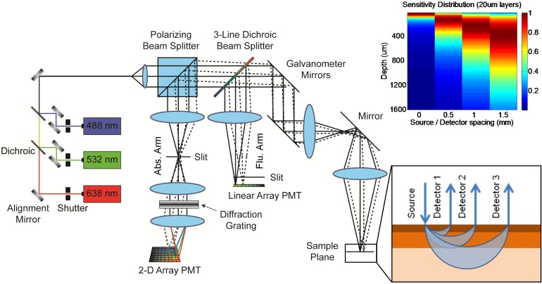
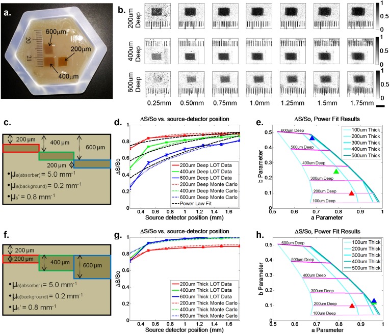
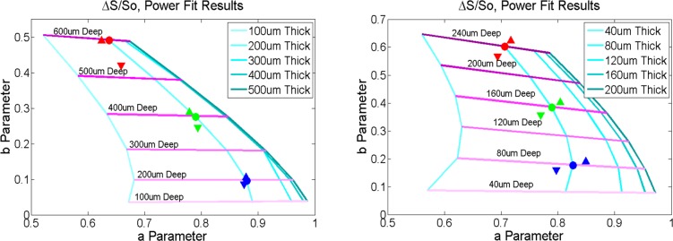
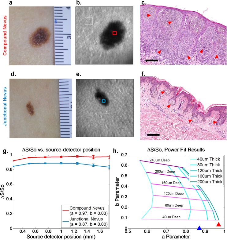
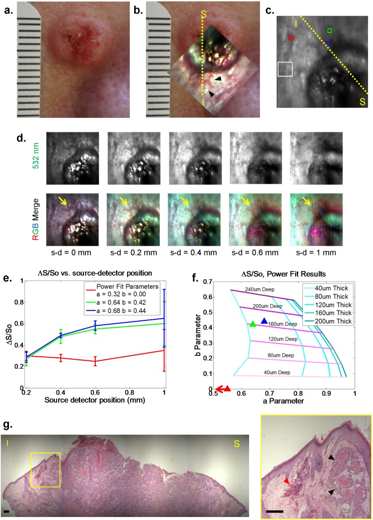
Evaluation of suspicious skin lesions by dermatologists is usually accomplished using white light examination and direct punch or surgical biopsy. However, these techniques can be imprecise for estimating a lesion's margin or level of dermal invasion when planning surgical resection. Laminar optical tomography (LOT) is an imaging technique capable of acquiring depth-sensitive information within scattering tissues. Here, we explore whether LOT data can be used to predict the depth and thickness of pigmented lesions using a range of simulations and phantom models. We then compare these results to LOT data acquired on normal and malignant skin lesions in vivo.
Keywords: (170.3880) Medical and biological imaging; (170.3890) Medical optics instrumentation.
Figures
References
-
- N. A. Howlader, N. M. Krapcho, N. Neyman, R. Aminou, S. F. Altekruse, C. L. Kosary, J. Ruhl, Z. Tatalovich, H. Cho, A. Mariotto, M. P. Eisner, D. R. Lewis, H. S. Chen, E. J. Feuer, and K. A. Cronin, eds., SEER Cancer Statistics Review, 1975-2009 (Vintage 2009 Populations), (National Cancer Institute, Bethesda, MD, 2012).
Grants and funding
LinkOut - more resources
Full Text Sources
Other Literature Sources