Review
. 2012:14:295-323.
doi: 10.1146/annurev-bioeng-071811-150114.

Regulation of cell behavior and tissue patterning by bioelectrical signals: challenges and opportunities for biomedical engineering

Affiliations
Review

Regulation of cell behavior and tissue patterning by bioelectrical signals: challenges and opportunities for biomedical engineering

Michael Levin et al. Annu Rev Biomed Eng. 2012.

Abstract

Achieving control over cell behavior and pattern formation requires molecular-level understanding of regulatory mechanisms. Alongside transcriptional networks and biochemical gradients, there functions an important system of cellular communication and control: transmembrane voltage gradients (V(mem)). Bioelectrical signals encoded in spatiotemporal changes of V(mem) control cell proliferation, migration, and differentiation. Moreover, endogenous bioelectrical gradients serve as instructive cues mediating anatomical polarity and other organ-level aspects of morphogenesis. In the past decade, significant advances in molecular physiology have enabled the development of new genetic and biophysical tools for the investigation and functional manipulation of bioelectric cues. Recent data implicate V(mem) as a crucial epigenetic regulator of patterning events in embryogenesis, regeneration, and cancer. We review new conceptual and methodological developments in this fascinating field. Bioelectricity offers a novel way of quantitatively understanding regulation of growth and form in vivo, and it reveals tractable, powerful control points that will enable truly transformative applications in bioengineering, regenerative medicine, and synthetic biology.


Figures

Figure 1
Figure 1
Membrane voltage is a key parameter regulating cell properties. A small sample of a much larger data set (taken after Reference 132) reveals the striking partitioning of cell types along the depolarized → polarized axis. Cells that are highly plastic (able to proliferate rapidly, undifferentiated) tend to be depolarized. Cells that are mature, terminally differentiated, and quiescent tend to be hyperpolarized. The mammalian liver is an interesting example—an adult tissue that exists close to the depolarized range and has unique regeneration potential. Importantly, Vmem is not simply a reflection of cell state but an instructive parameter: Artificial depolarization can confer neoplastic-like properties on somatic cells and prevent stem-cell differentiation, whereas artificial hyperpolarization can induce differentiation and suppress proliferation. Abbreviations: CHO, Chinese hamster ovary; hMSC, human mesenchymal stem cell; mESC, mouse embryonic stem cell.
Figure 2
Figure 2
Vmem gradients can be imaged in vivo. Voltage-sensitive fluorescent dyes (66) reveal a bioelectric map within complex tissues in vivo. This can be used to profile noninvasively the physiology of the tadpole tail during regenerative and nonregenerative conditions (a) (green indicates depolarization), the assembly of the tadpole face (b) (from Reference 87) (white arrows indicate hyperpolarized cell groups), and early embryogenesis (c) (frog embryo; red indicates depolarization, whereas blue indicates hyperpolarization).
Figure 3
Figure 3
Bioelectrical and genetic pathways form a cyclical dynamical system. The continuous interplay between biophysical and genetic mechanisms form a dynamical system: ion flows both control and are controlled by biochemical signals. Transcriptional events set up the expression of ion transporters, which regulate each other’s activity through physiological (posttranslational) mechanisms such as voltage gating of K+ channels. Transduction mechanisms (e.g., voltage-dependent regulation of entry of small signaling molecules through gap junctions) convert these signals into changes of gene expression. Changes in cell behavior and patterning can be driven not only by the well-known genetic cascades, but also directly by bioelectric cues that do not require changes in transcription or translation (such as movement and alignment of cells in electric fields as well as voltage-controlled movement of small signaling molecules through cell-membrane transporters).
Figure 4
Figure 4
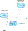
The state-space hypothesis of the bioelectric code. Membrane voltage is a powerful determinant of cellular state (94, 229), but a single parameter such as Vmem (Figure 1) is likely to be only a primitive approximation to the true richness of bioelectric control. Cell and tissue properties can be localized within a multidimensional physiological state space containing a number of orthogonal dimensions indicating membrane voltage, intracellular pH, K+ content, nuclear potential, Cl content, surface charge, etc. One hypothesis is that cells are grouped in distinct regions of this state space, corresponding to stem cells, tumor cells, somatic cells, and other interesting categories. This hypothesis implies that, given the necessary quantitative data, it will be possible to drive the desired changes in cell behavior (using pharmacologically targeting native channels/pumps and misexpression of well-characterized channel/pump constructs) by moving cell states into desired regions of this state space. For example, some cells may need to be depolarized by 20 mV and their internal pH acidified, to induce proliferation.
Figure 5
Figure 5
A model system for comprehensive physiomic profiling. The physiomic data set needed to flesh out the state-space hypothesis (Figure 4) requires high-resolution physiological data on multiple cell types from different organs and disease conditions. The merger of existing x,y,z,t,g (real-time three-dimensional anatomy and gene expression) data sets with physiological measurements will require that researchers develop transgenic model systems in which any desired cell/tissue of interest can be imaged in vivo. For example, transgenic frogs expressing the voltage reporter protein VSFP2.3 (schematized in panel a); (b) cell surface expression will allow microscopy approaches (c) to observe transmembrane potential data as a fluorescence resonance energy transfer (FRET) signal (d) in any tissue/organ/stage of interest, such as the sample data on the gradients among cells during embryonic development (e). Red arrow indicates a depolarized region of the cell membrane; yellow arrow indicates a polarized region of the cell membrane.
Figure 6
Figure 6
A model system for functional experiments in bioelectricity. (a) A vector can carry an insert encoding a depolarizing channel such as channelrhodopsin (ChR2) and one encoding a hyperpolarizing channel such as halorhodopsin, separated by the viral 2A sequence, thus allowing both proteins to be made from the same mRNA. Such protein variants (b) allow cells to be depolarized or hyperpolarized by exposure to light of a specific wavelength. Optical stimulation can be achieved by standard laser microscopy or special LEDs arranged in an array (c) that can, thus, provide patterned (tightly controlled with respect to the spatiotemporal pattern of imposed Vmem) control of the voltage gradient in any cell or tissue of a transgenic animal that constitutively expresses such a construct (d).

Similar articles

Cited by

References

    1. Robinson KR. 1989. Endogenous and applied electrical currents: their measurement and application. See Reference 26, pp. 1–25
    1. McCaig CD, Rajnicek AM, Song B, Zhao M. 2005. Controlling cell behavior electrically: current views and future potential. Physiol. Rev 85:943–78 - PubMed
    1. Fitzharris G, Baltz JM. 2006. Granulosa cells regulate intracellular pH of the murine growing oocyte via gap junctions: development of independent homeostasis during oocyte growth. Development 133:591–99 - PubMed
    1. Guthrie PB, Lee RE, Rehder V, Schmidt MF, Kater SB. 1994. Self-recognition: a constraint on the formation of electrical coupling in neurons. J. Neurosci 14:1477–85 - PMC - PubMed
    1. Nuccitelli R 2003. A role for endogenous electric fields in wound healing. Curr. Top. Dev. Biol 58:1–26 - PubMed

Publication types

LinkOut - more resources