. 2012 Jul 18;2(1):39.
doi: 10.1186/2191-219X-2-39.

Emerging role of radiolabeled nanoparticles as an effective diagnostic technique

Affiliations

Emerging role of radiolabeled nanoparticles as an effective diagnostic technique

Andréluís Branco de Barros et al. EJNMMI Res. .

Abstract

Nanomedicine is emerging as a promising approach for diagnostic applications. Nanoparticles are structures in the nanometer size range, which can present different shapes, compositions, charges, surface modifications, in vitro and in vivo stabilities, and in vivo performances. Nanoparticles can be made of materials of diverse chemical nature, the most common being metals, metal oxides, silicates, polymers, carbon, lipids, and biomolecules. Nanoparticles exist in various morphologies, such as spheres, cylinders, platelets, and tubes. Radiolabeled nanoparticles represent a new class of agent with great potential for clinical applications. This is partly due to their long blood circulation time and plasma stability. In addition, because of the high sensitivity of imaging with radiolabeled compounds, their use has promise of achieving accurate and early diagnosis. This review article focuses on the application of radiolabeled nanoparticles in detecting diseases such as cancer and cardiovascular diseases and also presents an overview about the formulation, stability, and biological properties of the nanoparticles used for diagnostic purposes.
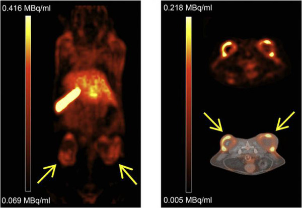
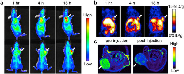
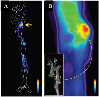
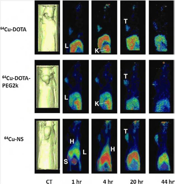

Figures

Figure 1
Figure 1
Passive versus active targeting. (Left) In passive targeting, particles tend to passively diffuse through the leaky vasculature of the tumor bed and accumulate primarily through the enhanced permeability effect. (Right) In active targeting, once particles have extravasated in the target tissue, the presence of ligands on the particle surface facilitates their interaction with receptors that are present on tumor or other cells, resulting in enhanced accumulation and preferential cellular uptake through receptor-mediated processes. This approach can be used either for vascular targeting and/or tumor cell targeting purposes. Reproduced with permission from [17].
Figure 2
Figure 2
The most common nanoparticles reported for diagnostic purposes.
Figure 3
Figure 3
Images of mice injected with 111In-loaded liposomes. (A) SPECT and (C) ex vivo phosphor imaging showed no focal, aortic arch hot spots in ApoE −/− mice injected with the nIgG probe, whereas all ApoE −/− mice injected with the LOX-1 probe revealed hot spots in the aortic arch ((B) includes sagittal, coronal, and transverse planes), confirmed by (D) ex vivo phosphor imaging. Sudan IV staining demonstrated comparable plaque distribution pattern for the (E and F) two groups. Reproduced with permission from [47].
Figure 4
Figure 4
MicroSPECT/CT images of C26 tumor-bearing BALB/c mice following injection of 188 Re-BMEDA-liposomes or188 Re-BMEDA. (a) Images of mice at 1 and 4 h after i.v. injection of 188Re-BMEDA. (b) Images of mice at 1, 4, 24, 48, and 72 h after injection of 188Re-BMEDA-liposome. Reproduced with permission from [49].
Figure 5
Figure 5
PET/CT images of 64Cu liposome distribution in HT29 tumor-bearing mice. Tumors were implanted on the right and left flanks. Coronal PET image 24 h after injection (left). Axial PET image (right top) and axial PET/CT fusion (right bottom) images 24 h after injection. Adapted with permission from [50].
Figure 6
Figure 6
In vivo NIRF (a) and PET (b) images of mouse injected with iron oxide nanoparticles. Images were acquired 1, 4, and 18 h after injection. (c) MRI images acquired before and 18 h after injection. Reproduced with permission from [73].
Figure 7
Figure 7
Autoradiography and fluorescence reflectance image of the aorta. (A) Autoradiography at an aneurysm in the descending thoracic aorta (arrow). (B) Fluorescence reflectance image of the same aorta. Nuclear and optical imaging concordantly showed nanoparticle accumulation in the aneurysmatic vessel wall. Adapted with permission from [75].
Figure 8
Figure 8
Sagittal PET images of three rats. The images were acquired at 1, 4, 20, 44 h after injection of 64Cu-DOTA, 64Cu-DOTA-PEG2k, and radiolabeled gold nanoshell (64Cu-NS), respectively. Surface-rendered CT images depicting tumor location are also shown (H, heart; L, liver; K, kidney; T, tumor). Reproduced with permission from [83].
Figure 9
Figure 9
Transaxial (a) and coronal MicroSPECT/CT (b) images of an athymic mouse with a C6-induced tumor. The images were taken 1 h after 99mTc-labeled gold nanoparticles conjugated with c[RGDfk(C)] intravenous administration. Adapted with permission from [84].
Figure 10
Figure 10
Imaging after administration of 111 In-labeled annexin A5-CPM in untreated and treated mice. (A) Dual SPECT/CT and (B) near-infrared fluorescence optical imaging after administration of 111In-labeled annexin A5-CPM into EL4 lymphoma-bearing mice (untreated animals). (C) Dual SPECT/CT and (D) near-infrared fluorescence optical imaging of EL4 lymphoma apoptosis after injection of 111In-labeled annexin A5-CPM into treated mice. Adapted with permission from [95].
Figure 11
Figure 11
Representative PET/CT images of 66 Ga-labeled nanographenes in 4T1 tumor-bearing mice at 3 h after injection. Tumor site is indicated by arrowheads. Adapted with permission from [126].
Figure 12
Figure 12
Representative whole-body coronal microPET images. The images were taken 4 h after injection, demonstrating M21 (left, arrow), M21L (middle, arrow), and enhanced M21 tumor contrast at 24 h after injection (right, arrow). Adapted with permission from [159].

References

    1. Moghimi SM, Hunter AC, Murray JC. Nanomedicine: current status and future prospects. FASEB J. 2005;19:311–330. doi: 10.1096/fj.04-2747rev. - DOI - PubMed
    1. Mailander V, Landfester K. Interaction of nanoparitcles with cells. Biomacromolecules. 2009;10:2379–2400. doi: 10.1021/bm900266r. - DOI - PubMed
    1. Wagner V, Dullaart A, Bock A, Zweck A. The emerging nanomedicine landscape. Nat Biotechnol. 2006;24:1211–1217. doi: 10.1038/nbt1006-1211. - DOI - PubMed
    1. Kim BYS, Rutka JT, Chan WCW. Current concepts: nanomedicine. N Eng J Med. 2010;363:2434–2443. doi: 10.1056/NEJMra0912273. - DOI - PubMed
    1. Lewis DR, Kamisoglu K, York AW, Moghe PV. Polymer-based therapeutics: nanoassemblies and nanoparticles for management of atherosclerosis. WIREs Nanomed Nanobiotechnol. 2011;3:400–420. doi: 10.1002/wnan.145. - DOI - PMC - PubMed

LinkOut - more resources