

Isolated cutaneous metastasis of uterine leiomyosarcoma: case report and review of literature
- PMID: 22809451
- PMCID: PMC3443420
- DOI: 10.1186/1746-1596-7-85
Isolated cutaneous metastasis of uterine leiomyosarcoma: case report and review of literature
Abstract
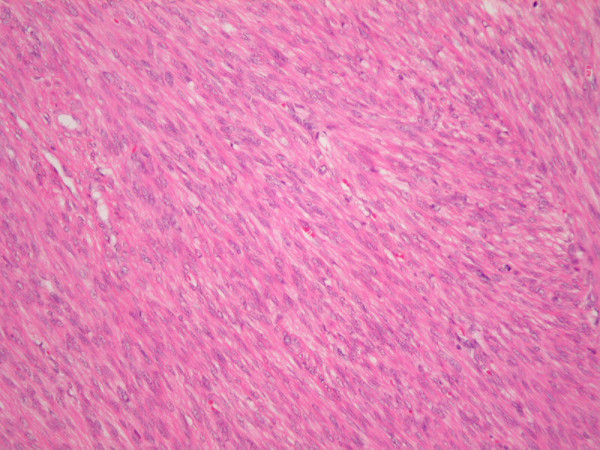
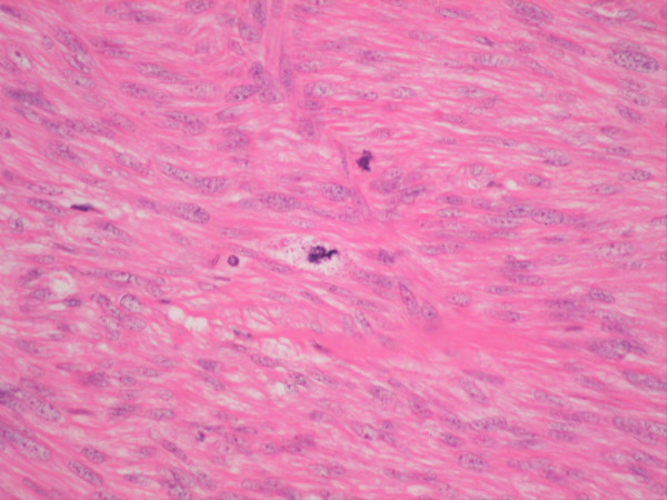
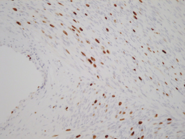
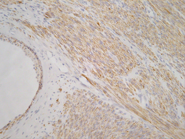
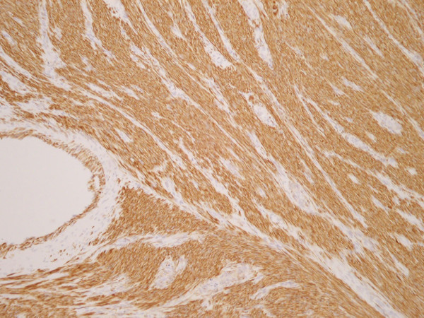
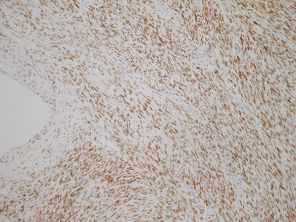
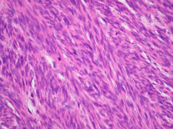
A 54 year old lady presented for routine excision of a scalp lesion thought clinically to represent a sebaceous cyst of the right occiput. 4 years earlier she underwent total abdominal hysterectomy and right salpingo-oophorectomy for 3 large uterine fibroids. Histo-pathological examination of the hysterectomy specimen revealed an incidental low-grade leiomyosarcoma. Staging imaging was negative for metastatic disease. She made an uneventful recovery and was treated further by adjuvant pelvic radiotherapy.She noticed an uncomfortable and unsightly cystic swelling on her occiput four years after hysterectomy and was referred for routine excision of what was believed to be a benign lesion. The lesion was excised and sent for histopathological examination. Microscopic analysis including immuno-histochemistry demonstrated an ER and PR positive metastatic deposit of leiomyosarcoma. The margins of excision were histologically clear of disease.At Multi-Disciplinary Team (MDT) discussion a diagnosis of metastatic scalp deposit from previous uterine leiomyosarcoma was made. Re-staging CT brain, thorax, abdomen and pelvis and MRI brain were negative for local recurrence or distant metastases. She is currently undergoing radiotherapy to the scalp and surrounding tissues and will be followed up closely by the involved teams.To the best of our knowledge, this is the first case described in the worldwide literature of isolated cutaneous metastasis to the scalp of uterine leiomyosarcoma without evidence of disseminated disease at other sites.
Virtual slides: The virtual slide(s) for this article can be found here: http://www.diagnosticpathology.diagnomx.eu/vs/1311834987345566.
Figures
References
-
- Golden T, Stout AP. Smooth muscle tumors of the gastrointestinal tract and retroperitoneal tissues. Surg Gynecol Obstet. 1941;73:784.
-
- Rolz-Cruz G, Kim CC. Tumor invasion of the skin. Dermatol Clin. 2008;26:89–102. - PubMed
Publication types
MeSH terms
LinkOut - more resources
Full Text Sources
Medical
Research Materials
