. 2012 Sep;23(17):3380-90.
doi: 10.1091/mbc.E12-02-0109. Epub 2012 Jul 18.

Cell cycle-regulated cortical dynein/dynactin promotes symmetric cell division by differential pole motion in anaphase

Affiliations

Cell cycle-regulated cortical dynein/dynactin promotes symmetric cell division by differential pole motion in anaphase

Elizabeth S Collins et al. Mol Biol Cell. 2012 Sep.

Abstract

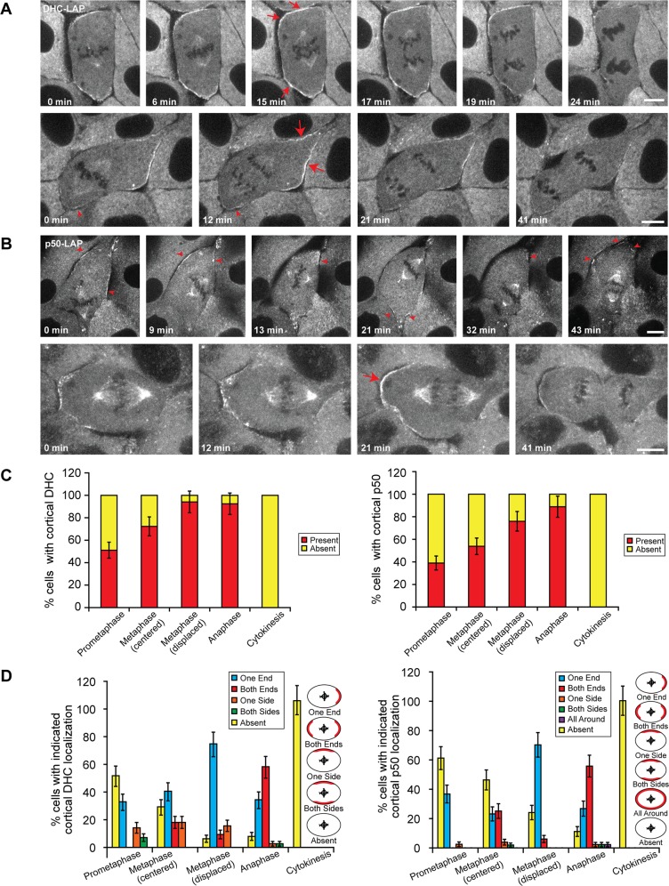
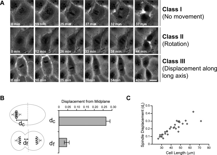
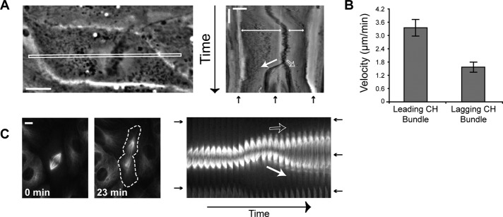
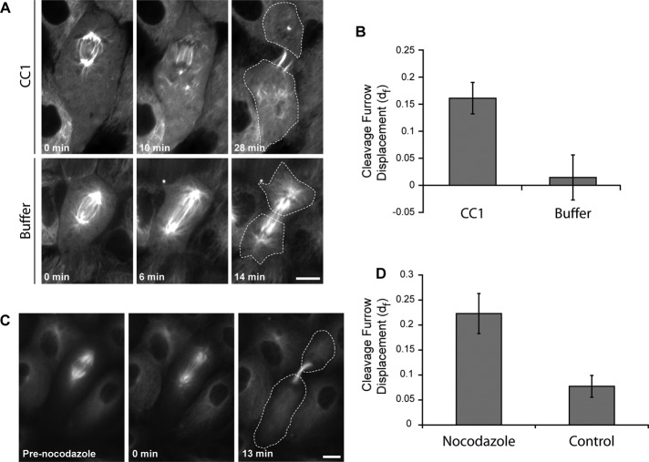
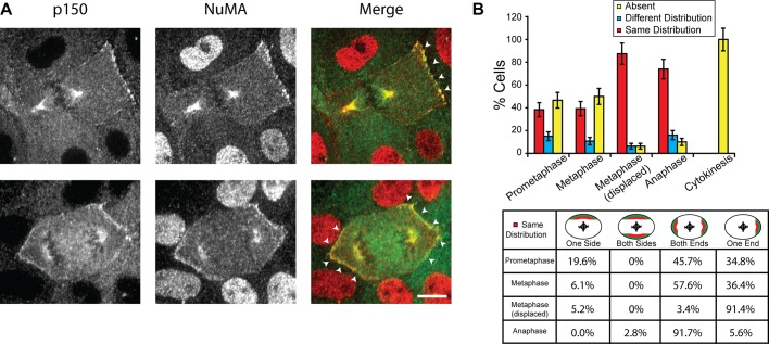
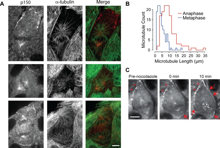
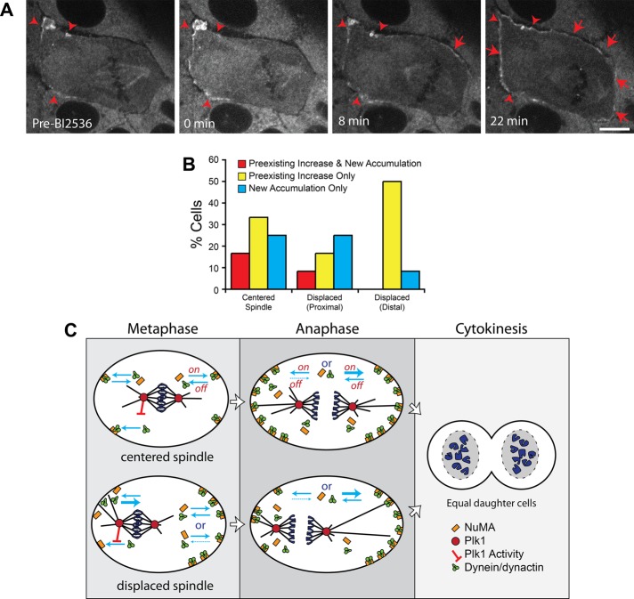
In cultured mammalian cells, how dynein/dynactin contributes to spindle positioning is poorly understood. To assess the role of cortical dynein/dynactin in this process, we generated mammalian cell lines expressing localization and affinity purification (LAP)-tagged dynein/dynactin subunits from bacterial artificial chromosomes and observed asymmetric cortical localization of dynein and dynactin during mitosis. In cells with asymmetrically positioned spindles, dynein and dynactin were both enriched at the cortex distal to the spindle. NuMA, an upstream targeting factor, localized asymmetrically along the cell cortex in a manner similar to dynein and dynactin. During spindle motion toward the distal cortex, dynein and dynactin were locally diminished and subsequently enriched at the new distal cortex. At anaphase onset, we observed a transient increase in cortical dynein, followed by a reduction in telophase. Spindle motion frequently resulted in cells entering anaphase with an asymmetrically positioned spindle. These cells gave rise to symmetric daughter cells by dynein-dependent differential spindle pole motion in anaphase. Our results demonstrate that cortical dynein and dynactin dynamically associate with the cell cortex in a cell cycle-regulated manner and are required to correct spindle mispositioning in LLC-Pk1 epithelial cells.


Figures

FIGURE 1:
FIGURE 1:
Dynamics of dynein and dynactin at the mitotic cortex of LLC-Pk1 cells. Time-lapse images of mitotic LLC-Pk1 cells expressing (A) DHC-LAP and (B) p50-LAP. (A) Top, a cell with centrally positioned spindle. Bottom, a cell with displaced mitotic spindle. (B) Top, a metaphase spindle becoming displaced before anaphase onset. Bottom, a cell with displaced mitotic spindle. Arrowheads, DHC-LAP signal at the proximal cortex (A, bottom) or dynamic p50-LAP signal (B, top). Arrows, enriched and uniform DHC-LAP and p50-LAP signal. Scale bars, 10 μm. (C, D) Quantitation of cortical dynein and dynactin localization throughout mitosis. (C) Percentage of cells exhibiting cortical dynactin and dynein localization. (D) Percentage of cells with indicated localization of cortical dynactin and dynein localization. Error bars in C and D represent SE of proportion; n = 167 (dynein) and 194 (dynactin) cells.
FIGURE 2:
FIGURE 2:
Classification of metaphase spindle movements in LLC-Pk1 cells. (A) Row 1, no spindle movement (class I). Row 2, rotational movement (class II). Row 3, displacement along long axis (class III). Scale bar, 10 μm. (B) Distance from the cell center to the chromosome plate measured at the last movie frame before anaphase (dc) and to the cleavage furrow at cytokinesis (df; p < 0.0001). Error bars represent SE of proportion. (C) Relationship between cell length along the interpolar axis and the normalized displacement of the spindle at anaphase onset. For A–C, n = 29 cells; for B and C, all classes of LLC-Pk1 cells were included in the measurement.
FIGURE 3:
FIGURE 3:
Asymmetric anaphase chromosome and pole movements result in symmetric division. (A) Phase contrast image of a cell with a displaced spindle (class III) at the start of time-lapse imaging and the corresponding kymograph of the boxed region. Scale bar, 10 μm (horizontal) and 10 min (vertical). (B) Average velocity of the leading and lagging chromosome bundles (n = 9 cells). Error bars, SE. (C) Differential pole movements during anaphase. GFP–α-tubulin images of a class III cell at t = 0 and 23 min and the corresponding kymograph. Dashed line outlines cell edge in the 23-min image. Scale bar, 10 μm. For A and C: closed white arrow, leading pole; open white arrow, lagging pole; double-headed arrow, spindle displacement; black arrow, cell edge and cleavage furrow.
FIGURE 4:
FIGURE 4:
Dynein activity is required for leading pole motion. (A) Class III cells expressing GFP–α-tubulin were injected with CC1 to inhibit dynein and dynactin interaction (top) or injected with buffer (bottom). Cells were microinjected immediately before t = 0 min. Dashed line outlines the cell edge in the final frame. Scale bar, 10 μm. (B) Cleavage furrow displacement in CC1- and buffer-injected cells (n = 10 and 6 cells, respectively). Error bars represent SE of proportion. p ≤ 0.01. (C) Time-lapse fluorescence images of an LLC-Pk1 cell expressing GFP–α-tubulin treated with 12.5 nM nocodazole. (D) Quantification of the cleavage furrow displacement in C for cells treated with 12.5 nM nocodazole or dimethyl sulfoxide control (n = 10 cells). p ≤ 0.01.
FIGURE 5:
FIGURE 5:
Codistribution of cortical NuMA and the p150 subunit of dynactin. (A) Immunofluorescence images of LLC-Pk1 cells stained with anti-p150 (left; green in merge) and anti-NuMA (middle; red in merge) antibodies; merged images to the right. Arrowheads, codistribution of NuMA and p150 at the cortex. Scale bar, 10 μm. (B) Top, quantitation of cortical NuMA and p150 distribution (n = 231 cells). Error bars represent SE of proportion. Bottom, patterns of NuMA and p150 codistribution observed in cells scored as same (red bars in B; n = 121 cells).
FIGURE 6:
FIGURE 6:
Dynein/dynactin accumulate at cortical sites distal to astral microtubules during metaphase. (A) Immunofluorescence images of LLC-Pk1 cells stained with anti-p150 antibodies (left; red in merge) and anti–α-tubulin (middle; green in merge) at metaphase with a displaced (top) or centered spindle (middle) and at anaphase with an elongated spindle and robust asters (bottom); merged images to the right. (B) Histograms of astral microtubule length during metaphase (blue) and anaphase (red; n = 30 cells; p < 0.0001). (C) Time-lapse fluorescence images of a metaphase cell expressing DHC-LAP treated with 33 μM nocodazole at t = 0 min. Arrowheads, preexisting cortical dynein patches; arrows, new dynein accumulation. Scale bars in A and C, 10 μm.
FIGURE 7:
FIGURE 7:
Inhibition of Plk1 kinase increases accumulation of cortical dynein. (A) Time-lapse fluorescence images of LLC-Pk1 cells expressing DHC-LAP arrested in metaphase with 5 μM MG132 and treated with 10 μM BI2536 at t = 0 min. Arrowheads, preexisting cortical dynein; arrows, new cortical dynein accumulation. Scale bar, 10 μm. (B) Quantitation of cortical dynein gain after BI2536 (n = 24 cells). (C) Model depicting cortical dynein in metaphase and anaphase cells. Top, centered spindles; bottom, displaced spindles. Plk1 inhibits the interaction between dynein/dynactin and cortical NuMA (red bar–headed line). Blue arrows represent on- and off-rates of dynein/dynactin and NuMA. In metaphase cells with a displaced spindle, the off-rate for dynein at the proximal cortex is increased (thick blue arrow). At the distal cortex, the on- and off-rates could be unchanged or the off-rate could be decreased relative to cells with a centered spindle. In anaphase, the on-rate is increased (thick blue arrow, right side) or, alternatively, the off-rate is decreased (dashed blue arrow, left side).

References

    1. Bruning-Richardson A, Langford KJ, Ruane P, Lee T, Askham JM, Morrison EE. EB1 is required for spindle symmetry in mammalian mitosis. PLoS One. 2011;6:e28884. - PMC - PubMed
    1. Burkhardt JK, Echeverri CJ, Nilsson T, Vallee RB. Overexpression of the dynamitin (p50) subunit of the dynactin complex disrupts dynein-dependent maintenance of membrane organelle distribution. J Cell Biol. 1997;139:469–484. - PMC - PubMed
    1. Busson S, Dujardin D, Moreau A, Dompierre J, De Mey JR. Dynein and dynactin are localized to astral microtubules and at cortical sites in mitotic epithelial cells. Curr Biol. 1998;8:541–544. - PubMed
    1. Carminati JL, Stearns T. Microtubules orient the mitotic spindle in yeast through dynein-dependent interactions with the cell cortex. J Cell Biol. 1997;138:629–641. - PMC - PubMed
    1. Cheeseman IM, Desai A. A combined approach for the localization and tandem affinity purification of protein complexes from metazoans. Sci STKE. 2005;2005:pl1. - PubMed

Publication types