. 2012 Sep;23(17):3370-9.
doi: 10.1091/mbc.E11-12-1010. Epub 2012 Jul 18.

Septin 7 forms a complex with CD2AP and nephrin and regulates glucose transporter trafficking

Affiliations

Septin 7 forms a complex with CD2AP and nephrin and regulates glucose transporter trafficking

Anita A Wasik et al. Mol Biol Cell. 2012 Sep.

Abstract

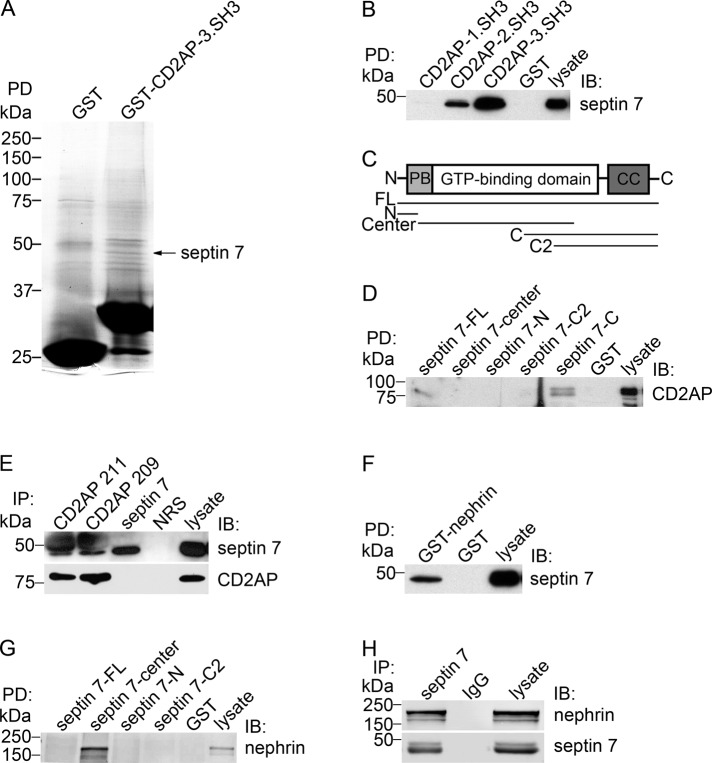
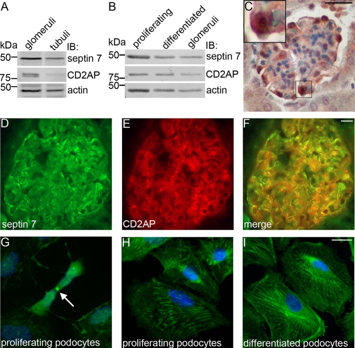
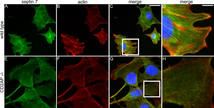
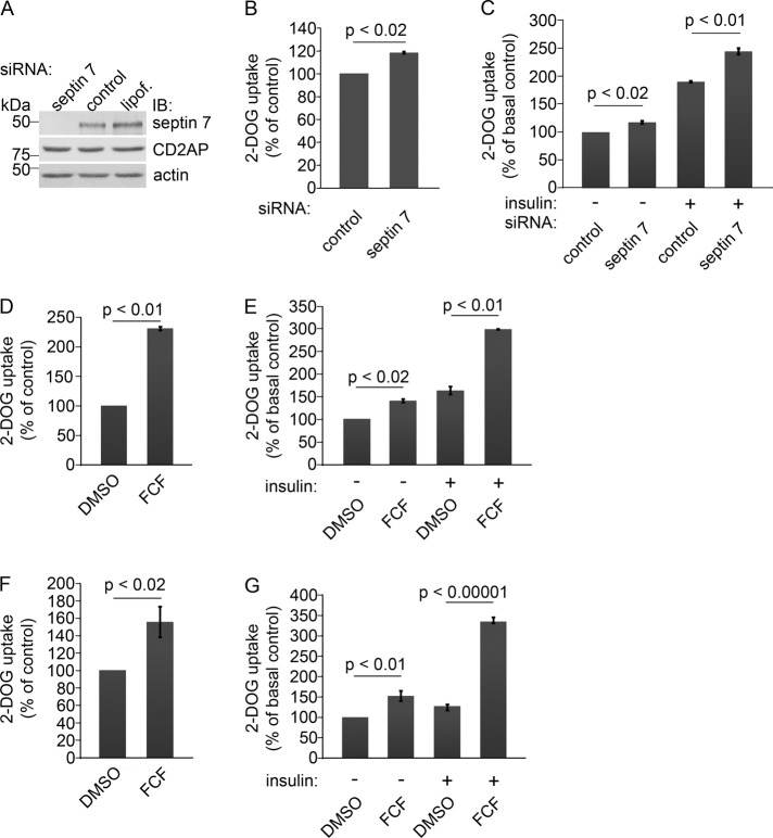
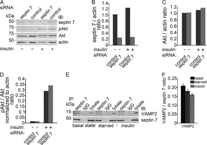
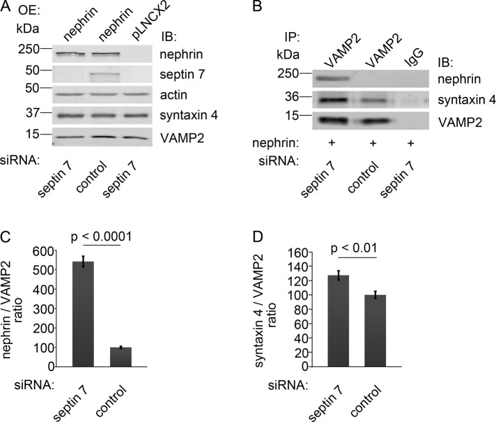
Podocytes are insulin-sensitive and take up glucose in response to insulin. This requires nephrin, which interacts with vesicle-associated membrane protein 2 (VAMP2) on GLUT4 storage vesicles (GSVs) and facilitates their fusion with the plasma membrane. In this paper, we show that the filament-forming GTPase septin 7 is expressed in podocytes and associates with CD2-associated protein (CD2AP) and nephrin, both essential for glomerular ultrafiltration. In addition, septin 7 coimmunoprecipitates with VAMP2. Subcellular fractionation of cultured podocytes revealed that septin 7 is found in both cytoplasmic and membrane fractions, and immunofluorescence microscopy showed that septin 7 is expressed in a filamentous pattern and is also found on vesicles and the plasma membrane. The filamentous localization of septin 7 depends on CD2AP and intact actin organization. A 2-deoxy-d-glucose uptake assay indicates that depletion of septin 7 by small interfering RNA or alteration of septin assembly by forchlorfenuron facilitates glucose uptake into cells and further, knockdown of septin 7 increased the interaction of VAMP2 with nephrin and syntaxin 4. The data indicate that septin 7 hinders GSV trafficking and further, the interaction of septin 7 with nephrin in glomeruli suggests that septin 7 may participate in the regulation of glucose transport in podocytes.


Figures

FIGURE 1:
FIGURE 1:
Septin 7 is a novel interaction partner of CD2AP and nephrin. (A) GST-fused 3.SH3 domain of CD2AP or GST alone were incubated with rat glomerular lysate, and the bound proteins were separated by SDS–PAGE, which was followed by staining with GelCode blue. The <50-kDa band detected in the CD2AP-GST pulldown was identified as septin 7 by mass spectrometry. (B) Septin 7 is pulled down with GST-2.SH3 and 3.SH3 domains of CD2AP, but not with GST alone, from rat glomerular lysate. (C) Schematic structure of septin 7 and GST-septin 7 fusion protein constructs used for pulldown assays. PB, polybasic region; CC, coiled-coil domain; FL, full-length (aa 1–418); N, NH2 terminus (aa 1–31); Center, central region (aa 32–298); C, COOH terminus (aa 179–418), and C2, COOH terminus 2 (aa 244–418). (D) Pulldown assays with GST-septin 7 fusion proteins covering full-length septin 7 (septin 7-FL), septin 7-center, septin 7 NH2 terminus (septin 7-N), and septin 7 COOH-terminal fragments (septin 7-C2 and septin 7-C) show that full-length septin 7 and the COOH terminus of septin 7 (septin-7-C) pull down CD2AP from rat glomerular lysate. (E) Two different CD2AP antibodies (CD2AP 211 and 209) coimmunoprecipitate endogenous septin 7 in HEK293 cells. Immunoprecipitation with septin 7 antibodies indicates septin 7 band immediately below the IgG heavy chain. Neither septin 7 nor CD2AP are present in immunoprecipitations with normal rabbit serum (NRS). (F) GST-nephrin cytoplasmic domain, but not GST alone, pulls down septin 7 from rat glomerular lysate. (G) Pulldown assays with GST-septin 7 fusion proteins indicate that the central domain of septin 7, but not GST alone, pulls down nephrin from rat glomerular lysate. (H) Nephrin coimmunoprecipitates with septin 7 antibodies, but not with rabbit IgG from rat glomerular lysate. Isolated glomeruli or HEK293 cells were lysed in 1% NP-40, 20 mM HEPES (pH 7.5), and 150 mM NaCl, incubated with CD2AP, septin 7, or nephrin GST-fusion proteins or antibodies, and the precipitated proteins were immunoblotted with anti-CD2AP, anti–septin 7, or anti-nephrin IgG. Glomerular or HEK293 cell lysates (10 μg) are included as control.
FIGURE 2:
FIGURE 2:
Septin 7 is expressed in podocytes. (A) Immunoblotting of rat glomerular and tubular fractions shows septin 7 expression in glomeruli and tubuli and CD2AP expression in glomeruli. (B) Septin 7 and CD2AP are expressed in both proliferating and differentiated cultured human podocytes. (C) Mouse kidney section stained with septin 7 antibody shows septin 7 expression in glomeruli, where it localizes in podocytes. Higher magnification of the boxed region is shown in the inset. Septin 7 (D) partially colocalizes with CD2AP (E) in podocytes in rat kidney sections as visualized in the merged image (F). (G) Septin 7 localizes in midbody (arrow) in proliferating podocytes and is observed in filamentous and punctate pattern in both proliferating (H) and differentiated (I) podocytes. In (A) and (B), rat glomerular and tubular fractions and cultured human podocytes were lysed in 1% NP-40, 20 mM HEPES (pH 7.5), and 150 mM NaCl, and immunoblotted with septin 7 and CD2AP IgGs and actin IgG as a loading control. (C) Adult mouse kidney paraffin sections were processed for immunoperoxidase staining, labeled with septin 7 IgG, and analyzed by light microscopy. In (D–F) and (G–I), rat kidney sections and cultured human podocytes were fixed with acetone and paraformaldehyde (PFA), respectively; labeled with septin 7 and CD2AP IgG; and examined by immunofluorescence. Scale bar: 20 μm.
FIGURE 3:
FIGURE 3:
Localization of septin 7 depends on CD2AP. Double labeling of wild-type (A–D) and CD2AP−/− (E–H) mouse podocytes for septin 7 (green) and actin (red). Nuclei are visualized with Hoechst (blue). (A–D) In wild-type podocytes, septin 7 filaments (A) partially align along actin stress fibers detected with phalloidin (B–D). (E–H) In CD2AP−/− podocytes, septin 7 filaments disappear. Boxed regions indicated in (C and G) are magnified in (D and H), respectively. Cultured mouse podocytes were fixed with PFA, labeled with septin 7 IgG and phalloidin, and examined by fluorescence microscopy. Scale bars: 20 µm (A–C and E–G); 7 µm (D and H).
FIGURE 4:
FIGURE 4:
Knockdown of septin 7 or alteration of the assembly of septins increases glucose uptake in HIRc cells and podocytes. (A) Septin 7 siRNA leads to 81% reduction in septin 7 expression in HIRc cells. CD2AP expression level remains unchanged in septin 7 knockdown cells. HIRc cells were transfected with rat septin 7 SMARTpool siRNA (septin 7), siCONTROL Non-Targeting Pool siRNA (control), or Lipofectamine 2000 alone (lipof.), and analyzed by immunoblotting 72 h after transfection. Actin is included as a loading control. (B) Depletion of septin 7 increases the glucose uptake activity of HIRc cells by 19% compared with the control siRNA-transfected cells (set to 100%) in basal state. (C) Knockdown of septin 7 increases the glucose uptake activity of HIRc cells by 17% in serum-starved cells (septin 7 siRNA, − insulin) and by 144% after insulin stimulation (septin 7 siRNA, + insulin). The increase in glucose uptake in control siRNA-transfected cells is 90% after insulin stimulation (control siRNA, + insulin). Glucose uptake activity of the control siRNA-transfected and serum-starved cells is set to 100% (control siRNA, − insulin). (D) HIRc cells treated with FCF show 131% increase in glucose uptake activity in basal state compared with solvent only–treated cells (DMSO, set to 100%). (E) FCF treatment increases the glucose uptake activity of HIRc cells by 41% in serum-starved cells (FCF, − insulin) and by 198% after insulin stimulation (FCF, + insulin). The increase in glucose uptake in solvent only–treated cells is 64% after insulin stimulation (DMSO, + insulin). Glucose uptake activity of the solvent only–treated and serum-starved cells is set to 100% (DMSO, − insulin). (F) Alteration of septin assembly by FCF in mouse podocytes leads to a 56% increase in glucose uptake in basal state. (G) FCF treatment increases the glucose uptake activity of mouse podocytes by 52% in serum-starved cells (FCF, − insulin) and by 235% after insulin stimulation (FCF, + insulin). Solvent only–treated mouse podocytes show a 27% increase in glucose uptake after insulin stimulation (DMSO, + insulin). HIRC cells were transfected with septin-7 siRNA (A–C) or treated with 50 μM FCF (D and E), and glucose uptake was measured as described in Materials and Methods in basal state (B and D) or after serum starvation (−) and treatment with 200 nM insulin (+) (C and E). Mouse podocytes were treated with 50 μM FCF for 4 h (F and G) and treated (+) or not (−) with 20 nM insulin (G). Bars show the mean and error bars show the SD of three independent experiments using Student's t test.
FIGURE 5:
FIGURE 5:
Septin 7 forms a complex with VAMP2 involved in vesicle trafficking. (A) Knockdown of septin 7 does not affect the activity of insulin-mediated Akt signaling pathway. HIRc cells transfected with septin 7 or control siRNA were serum-starved and untreated (−) or treated (+) with 200 nM insulin and immunoblotted with septin 7, pan-Akt (Akt), phospho-Akt (pAkt), and actin IgG. Akt phosphorylation is unaffected by septin 7 depletion. Representative blot of three independent experiments. (B) Quantification of septin 7 level in (A) presented as septin 7:actin ratio. (C) Quantification of total Akt level in (A) presented as Akt:actin ratio. (D) Quantification of pAkt levels in (A) presented as pAkt:total Akt normalized to actin ratio. (E) VAMP2 coimmunoprecipitates with septin 7 but not with control IgG in HIRc cells in basal state and in serum-starved and insulin-stimulated cells. Lysate, 10 μg. (F) Quantification of protein levels of three replicate blots as in (E) presented as VAMP2:septin 7 ratio showing that insulin stimulation does not affect the interactions as the difference in complex formation between different conditions did not reach statistical significance.
FIGURE 6:
FIGURE 6:
Knockdown of septin 7 increases complex formation between nephrin and VAMP2, and between syntaxin 4 and VAMP2. (A) HIRc cells were transfected with septin 7 or control siRNA, which was followed by nephrin or empty vector (pLNCX2) overexpression (OE) by viral infection. Immunoblotting confirms nephrin overexpression and septin 7 depletion. Syntaxin 4 and VAMP2 levels remain constant. (B) Cell lysates of HIRc cells, transfected with septin 7 or control siRNA and overexpressing nephrin, were immunoprecipitated with anti-VAMP2 antibodies and immunoblotted with nephrin, syntaxin 4, and VAMP2 antibodies. Depletion of septin 7 increases the interaction of VAMP2 with nephrin and syntaxin 4. (C and D) Quantification of protein levels of three replicate blots as in (B). The bars represent nephrin:VAMP2 (C) and syntaxin 4:VAMP2 (D) ratios. Bars show the mean and error bars show the SD using Student's t test.

Similar articles

Cited by

References

    1. Ahola H, Heikkila E, Astrom E, Inagaki M, Izawa I, Pavenstadt H, Kerjaschki D, Holthofer H. A novel protein, densin, expressed by glomerular podocytes. J Am Soc Nephrol. 2003;14:1731–1737. - PubMed
    1. Beites CL, Campbell KA, Trimble WS. The septin Sept5/CDCrel-1 competes with α-SNAP for binding to the SNARE complex. Biochem J. 2005;385:347–353. - PMC - PubMed
    1. Beites CL, Xie H, Bowser R, Trimble WS. The septin CDCrel-1 binds syntaxin and inhibits exocytosis. Nat Neurosci. 1999;2:434–439. - PubMed
    1. Cormont M, Meton I, Mari M, Monzo P, Keslair F, Gaskin C, McGraw TE, Le Marchand-Brustel Y. CD2AP/CMS regulates endosome morphology and traffic to the degradative pathway through its interaction with Rab4 and c-Cbl. Traffic. 2003;4:97–112. - PubMed
    1. Coward RJ, Welsh GI, Koziell A, Hussain S, Lennon R, Ni L, Tavare JM, Mathieson PW, Saleem MA. Nephrin is critical for the action of insulin on human glomerular podocytes. Diabetes. 2007;56:1127–1135. - PubMed

Publication types

MeSH terms

LinkOut - more resources