Hip capsule dimensions in patients with femoroacetabular impingement: a pilot study
- PMID: 22810156
- PMCID: PMC3492636
- DOI: 10.1007/s11999-012-2485-2
Hip capsule dimensions in patients with femoroacetabular impingement: a pilot study
Abstract
Background: Joint-preserving hip surgery, either arthroscopic or open, increasingly is used for the treatment of symptomatic femoroacetabular impingement (FAI). As a consequence of surgery, thickening of the joint capsule and intraarticular adhesions between the labrum and joint capsule and between the femoral neck and the joint capsule have been observed. These alterations are believed to cause persistent pain and reduced range of motion. Because the diagnosis is made with MR arthrography, knowledge of the normal capsular anatomy and thickness on MRI in patients is important. To date there is no such information available.
Questions/purposes: The purpose of this study was to establish thickness, length of the hip capsule, and the size of the perilabral recess in patients with FAI.
Methods: We reviewed the preoperative MR arthrography of 30 patients (15 men) with clinical symptoms of FAI. We measured capsular thickness and made observations on the perilabral recess.
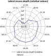
Results: The joint capsule was thickest (6 mm) anterosuperiorly between 1 and 2 o'clock. The average length from the femoral head-neck junction to the femoral insertion of the capsule ranged from 19 to 33 mm. A perilabral recess was present circumferentially, even across the acetabular notch, where the labrum is supported by the transverse acetabular ligament. The shortest recess occurred superiorly.
Conclusions: Knowledge of the capsular anatomy in patients with FAI before surgery is important to judge the postoperative changes and to plan potential further therapy including arthroscopic treatment of intraarticular adhesions.
Figures


Similar articles
-
Hip capsular thickness correlates with range of motion limitations in femoroacetabular impingement.Knee Surg Sports Traumatol Arthrosc. 2018 Oct;26(10):3178-3187. doi: 10.1007/s00167-018-4915-5. Epub 2018 Mar 24. Knee Surg Sports Traumatol Arthrosc. 2018. PMID: 29574547
-
Capsular attachment on the anterosuperior femoral head-neck junction: A hypothesis about femoroacetabular impingement.J Anat. 2024 Aug;245(2):231-239. doi: 10.1111/joa.14046. Epub 2024 Apr 8. J Anat. 2024. PMID: 38590168 Free PMC article.
-
Real-Time Assessment of Femoroacetabular Motion Using Radial Gradient Echo Magnetic Resonance Arthrography at 3 Tesla in Routine Clinical Practice: A Pilot Study.Arthroscopy. 2019 Aug;35(8):2366-2374. doi: 10.1016/j.arthro.2019.02.049. Arthroscopy. 2019. PMID: 31395172
-
Postoperative imaging in femoroacetabular impingement.Semin Musculoskelet Radiol. 2013 Jul;17(3):272-8. doi: 10.1055/s-0033-1348093. Epub 2013 Jun 20. Semin Musculoskelet Radiol. 2013. PMID: 23787981 Review.
-
Groin pain after open FAI surgery: the role of intraarticular adhesions.Clin Orthop Relat Res. 2009 Mar;467(3):769-74. doi: 10.1007/s11999-008-0653-1. Epub 2008 Dec 10. Clin Orthop Relat Res. 2009. PMID: 19082679 Free PMC article. Review.
Cited by
-
Correlation of hip capsule morphology with patient symptoms from femoroacetabular impingement.J Orthop Res. 2021 Mar;39(3):590-596. doi: 10.1002/jor.24788. Epub 2020 Jul 6. J Orthop Res. 2021. PMID: 32592526 Free PMC article.
-
Quantification and correlation of hip capsular volume to demographic and radiographic predictors.Knee Surg Sports Traumatol Arthrosc. 2016 Jun;24(6):2009-15. doi: 10.1007/s00167-014-3275-z. Epub 2014 Sep 14. Knee Surg Sports Traumatol Arthrosc. 2016. PMID: 25218574
-
The envelope of passive motion allowed by the capsular ligaments of the hip.J Biomech. 2015 Nov 5;48(14):3803-9. doi: 10.1016/j.jbiomech.2015.09.002. Epub 2015 Sep 14. J Biomech. 2015. PMID: 26429769 Free PMC article.
-
A quantitative analysis of hip capsular thickness.Knee Surg Sports Traumatol Arthrosc. 2015 Sep;23(9):2548-53. doi: 10.1007/s00167-014-3030-5. Epub 2014 May 10. Knee Surg Sports Traumatol Arthrosc. 2015. PMID: 24817105
-
Normal anatomical variants of the labrum of the hip at magnetic resonance imaging: a systematic review.Eur Radiol. 2013 Jun;23(6):1694-710. doi: 10.1007/s00330-012-2744-3. Epub 2012 Dec 18. Eur Radiol. 2013. PMID: 23247809
References
MeSH terms
LinkOut - more resources
Full Text Sources
Research Materials

