. 2012 Oct;40(18):9060-72.
doi: 10.1093/nar/gks674. Epub 2012 Jul 18.

Lesion-specific DNA-binding and repair activities of human O⁶-alkylguanine DNA alkyltransferase

Affiliations

Lesion-specific DNA-binding and repair activities of human O⁶-alkylguanine DNA alkyltransferase

Manana Melikishvili et al. Nucleic Acids Res. 2012 Oct.

Abstract

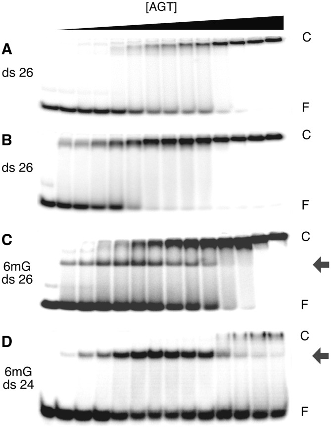
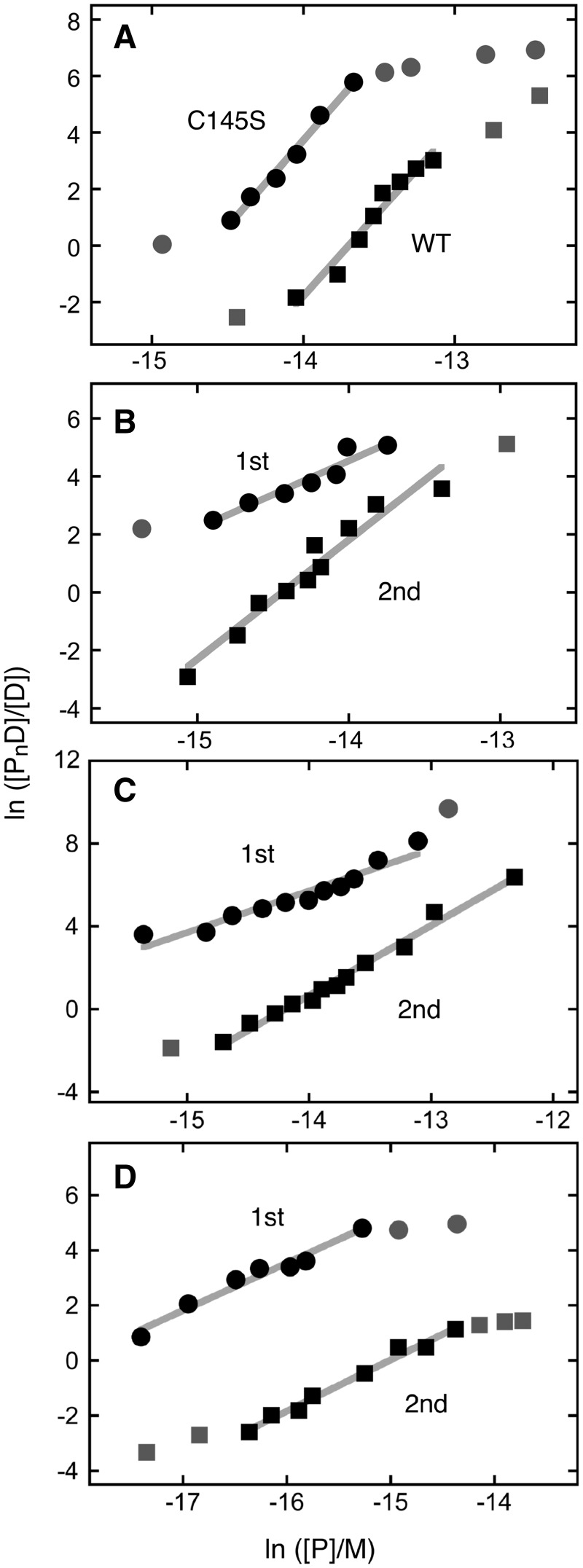
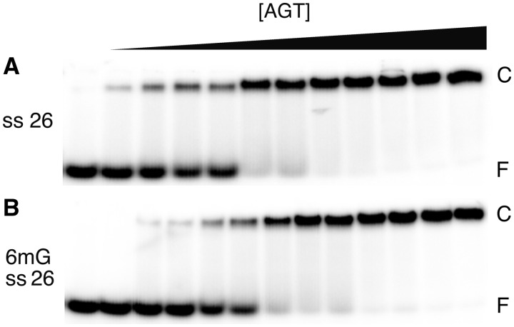
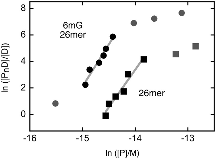
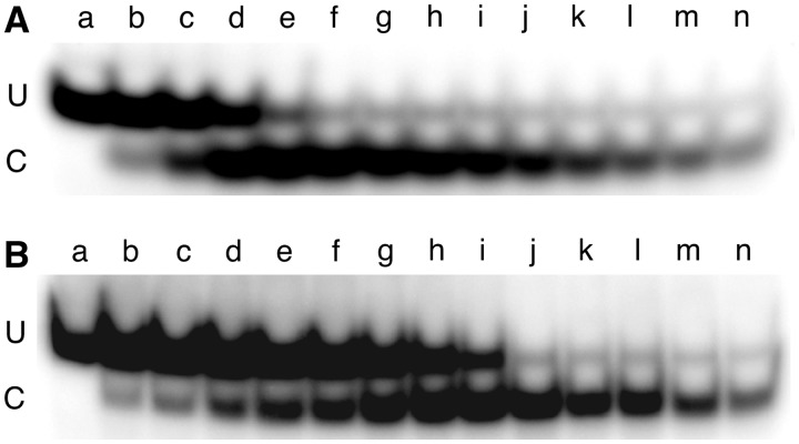
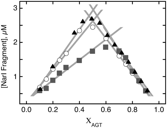
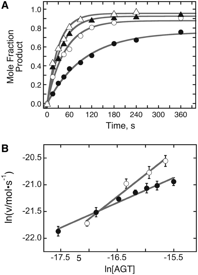
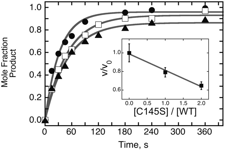
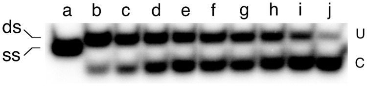
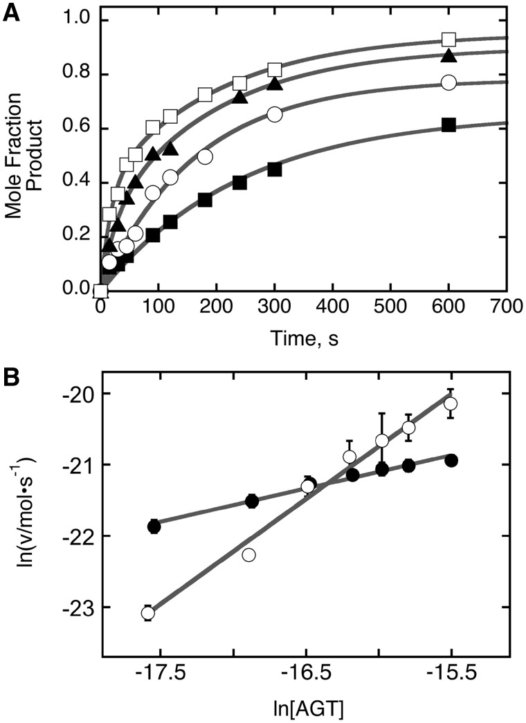
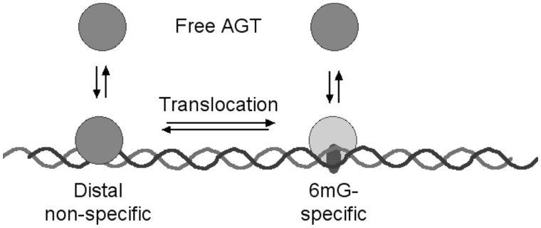
Binding experiments with alkyl-transfer-active and -inactive mutants of human O(6)-alkylguanine DNA alkyltransferase (AGT) show that it forms an O(6)-methylguanine (6mG)-specific complex on duplex DNA that is distinct from non-specific assemblies previously studied. Specific complexes with duplex DNA have a 2:1 stoichiometry that is formed without accumulation of a 1:1 intermediate. This establishes a role for cooperative interactions in lesion binding. Similar specific complexes could not be detected with single-stranded DNA. The small difference between specific and non-specific binding affinities strongly limits the roles that specific binding can play in the lesion search process. Alkyl-transfer kinetics with a single-stranded substrate indicate that two or more AGT monomers participate in the rate-limiting step, showing for the first time a functional link between cooperative binding and the repair reaction. Alkyl-transfer kinetics with a duplex substrate suggest that two pathways contribute to the formation of the specific 6mG-complex; one at least first order in AGT, we interpret as direct lesion binding. The second, independent of [AGT], is likely to include AGT transfer from distal sites to the lesion in a relatively slow unimolecular step. We propose that transfer between distal and lesion sites is a critical step in the repair process.


Figures

Figure 1.
Figure 1.
Discrete, high-mobility complexes form when AGT binds 6mG-containing dsDNAs. EMSAs performed at 20 ±1°C. (A) Titration of unmodified double stranded 26 bp DNA (oligo 1 + 3 duplex; 0.17 µM) with wild-type AGT (0–9.6 µM) to form the low-mobility cooperative complex. The equilibration buffer was 10 mM Tris (pH 8.3 at 20°C), 50 mM NaCl, 0.1 mM EDTA, 1 mM DTT, 5% glycerol, 0.1 mg/mL BSA (bovine serum albumin). Samples resolved in a 15% native polyacrylamide gel cast and run as described. (B) Titration of unmodified double stranded 26 bp DNA (oligo 1 + 3 duplex; 0.17 µM) with C145S-AGT (0–9.5 µM). Equilibration buffer and gel conditions as in (A). (C) Titration of 6mG-containing 26 bp duplex DNA (oligo 2 + 3 duplex; 0.15 µM) with C145S-AGT (0–5.1 µM). Equilibration buffer and gel conditions as in (A). (D) Titration of 6mG-containing 24 bp duplex DNA (oligo 4 + 5 duplex; 0.038 µM) with C145S-AGT (0–2.0 µM). Equilibration buffer was 40 mM Tris-acetate (pH 7.9 at 20°C), 100 mM potassium acetate, 20 mM magnesium acetate, 2 mM DTT, 0.5 mg/mL BSA. These samples resolved in a 20% native polyacrylamide gel cast and run as described. Band designations: F, free DNA; C, cooperative AGT-DNA complex; grey arrows, 6mG-specific complexes.
Figure 2.
Figure 2.
Graphs of the dependence of log[PnD]/[D] on log[P] for AGT-complexes with duplex DNAs. Data are from the experiments shown in Figure 1 and others that provide additional [AGT] values. The lines represent least-squares fits to the data ensembles for the ranges about the midpoint of each reaction, with [AGT]free calculated as described in Experimental Procedures. Symbols: the points used in the fit are indicated by the black symbols; other points in the data sets are indicated by grey symbols. (A) Data from titrations of unmodified double stranded 26 bp DNA (oligos 1 + 3) with C145S-AGT (filled circle; displaced upward 3 units for clarity) and wild-type AGT (filled square). The slopes are 5.97 ± 0.49 and 6.09 ± 0.32, respectively. (B) Data from titration of 6mG-containing double stranded 26 bp DNA (oligos 2 + 3) with alkyltransfer-active P140K-AGT. First binding step (filled circle; points displaced upward 4 units for clarity); second binding step (filled square). The slopes are 2.33 ± 0.29 and 4.11 ± 0.34, respectively. (C) Data from titration of 6mG-containing double stranded 26 bp DNA (oligos 2 + 3) with C145S-AGT. First binding step (filled circle; points displaced upward 5 units for clarity); second binding step (filled square). The slopes are 2.01 ± 0.19 and 3.37 ± 0.12, respectively. (D) Data from titration of 6mG-containing double stranded 24 bp DNA (oligos 4 + 5) with C145S-AGT. First binding step (filled circle; points displaced upward 3 units for clarity); second binding step (filled square). The slopes are 1.72 ± 0.14 and 1.86 ± 0.09, respectively.
Figure 3.
Figure 3.
Single-step binding to ssDNAs. EMSAs performed at 20 ± 1°C. (A) Titration of unmodified single stranded 26 nt DNA (oligo 1, 0.17 µM) with C145S-AGT (0–2.2 µM) to form the low-mobility cooperative complex. The equilibration buffer was 10 mM Tris (pH 8.3 at 20°C), 50 mM NaCl, 0.1 mM EDTA, 1 mM DTT, 5% glycerol, 0.1 mg/ml BSA. Samples resolved in 20% native polyacrylamide gels cast and run as described. (B) Titration of 6mG containing 26 nt DNA (oligo 2; 0.19 µM) with C145S-AGT (0–3.4 µM). The equilibration buffer and electrophoresis conditions were as in (A).
Figure 4.
Figure 4.
Graphs of the dependence of log[PnD]/[D] on log[P] for C145S-AGT-complexes with ssDNAs. Data are from the experiments shown in Figure 3. The lines represent least-squares fits to the data ensemble for the range about the midpoint of each reaction, with [AGT]free calculated as described in Experimental Procedures. Symbols: the points used in the fit are indicated by the black symbols; other points in the data sets are indicated by grey symbols. Titration of 6mG-containing 26mer (oligo 2, filled circle, displaced upward 4 units for clarity) and unmodified 26mer (oligo 1, filled square). The slopes are 6.35 ± 0.64 and 5.64 ± 0.62, respectively.
Figure 5.
Figure 5.
Continuous-variation NarI DNA repair assays. Images of 20% polyacrylamide gels resolving NarI cleaved fragment (C) from uncleaved DNA (U). The DNA used was the double-stranded 6mG-containing 24mer (oligos 5 + 6) and oligo 6 carried a 5′-32P label. The reaction buffer was 20 mM Tris acetate (pH 7.9), 50 mM potassium acetate, 10 mM magnesium acetate, 1 mM DTT. (A) DNA and wild-type AGT solutions (each 5.8 µM) were mixed in volume ratios giving mole-fractions of AGT ranging from 0.1 to 0.9. For samples a–n, these were 0, 0.1, 0.15, 0.2, 0.36, 0.42, 0.5, 0.55, 0.6, 0.66, 0.72, 0.82, 0.86 and 0.92, respectively. Incubation was for 3 h at 20 ± 0.1°C. Repair reactions were quenched by addition of SDS to a final concentration of 0.2%, and samples were extracted with phenol, then ether, as described, then digested with 10U of NarI for 2 h at 37°C, prior to electrophoresis. (B) Effect of mixing wild-type and C145S-AGTs in 1:1 molar ratio. Assays carried out as described above. For samples a–n, the mole fractions of AGT (wild-type plus C145A) were 0, 0.1, 0.15, 0.2, 0.25, 0.3, 0.4, 0.5, 0.6, 0.7, 0.75, 0.8, 0.85 and 0.9, respectively.
Figure 6.
Figure 6.
Continuous variation analyses of dsDNA-repair reactions. The data are from the experiments shown in Figure 5, plus an additional one carried out with P140K-AGT. Symbols: wild-type AGT (filled triangle); P140K mutant (open circle); wild-type- and C145S-AGTs in 1:1 ratio (filled square). The solid lines are linear fits to rising and falling subsets of the data. Optimal combining ratios are given by their intersection. For wild-type- and P140K-AGTs and the wild-type plus C145S-AGT mixture, these were 0.503, 0.51 and 0.65, respectively.
Figure 7.
Figure 7.
Time-dependence of dsDNA repair detected by NarI sensitivity. The 6mG-containing 24mer duplex (oligos 4 and 5) 0.037 µM, was dissolved in 40 mM Tris acetate (pH 7.9 at 20°C), 100 mM potassium phosphate, 20 mM magnesium acetate, 2 mM DTT, containing 0.5 mg/ml BSA. To start the reaction, AGT was added to a final concentration of 0.11 µM. Aliquots were withdrawn and quenched in 0.2% SDS after 15 s, 30 s, 45 s, 60 s, 120 s, 180 s, 240 s, 360 s, 900 s, 2400 s and 3600 s (samples b–l, respectively); unrepaired DNA is shown in sample a. Samples were deproteinized by phenol extraction followed by ether extractions as described; they were then digested with NarI (15 U) for 2 h at 37°C and resolved by electrophoresis on a 20% gel.
Figure 8.
Figure 8.
Analysis of repair reactions carried out on duplex DNA at several AGT concentrations. (A) Time profiles for repair reactions. Data were obtained as shown in Figure 7, with [6mG DNA] = 0.037 µM and [AGT] = 0.037 µM (filled circle); [AGT] = 0.074 µM (open square); [AGT] = 0.11 µM (filled triangle); [AGT] = 0.148 µM (open triangle). The smooth curves are fits of Equation (2) to the data. (B) Graphs of the dependence of ln v as functions of ln [AGT] for repair reactions run with [DNA] = 0.037 µM (open circle) and [DNA] = 0.023 µM (filled circle). Here v is the initial reaction rate; error bars correspond to the 95% confidence intervals on v determined from fits like those shown in Panel A. The solid lines are linear fits to the data as described for Equation (3); the slopes provide estimates of the order of the reaction in AGT. For [DNA] = 0.037 µM, α = 0.83 ± 0.08; for [DNA] = 0.023 µM α = 0.47 ± 0.03.
Figure 9.
Figure 9.
Effect of alkyl-transfer-inactive AGT on dsDNA repair. All samples contained 32P-labeled 6mG-duplex 24mer DNA (0.037 µM) and wild-type AGT (0.111 µM); samples contained, in addition, C145S-AGT at 0 µM (filled circle), 0.111 µM (open square) or 0.222 µM (filled triangle). The time course of repair was measured as described for Figures 7 and 8. Inset: graph of relative velocity in the presence of C145S-AGT (v) with respect to velocity in its absence (v0) as a function of the molar ratio [C145S]/[WT] in each assay. The solid line is a linear fit to the data.
Figure 10.
Figure 10.
Time course of repair for a ssDNA. The single-stranded 6mG-24mer (oligo 4) was 5′-labeled with 32P. Repair was carried out as described for Figure 7 in a solution containing 0.023 µM DNA and 0.092 µM AGT. Samples b–k correspond to reactions stopped by addition of 0.2% SDS after 15 s, 30 s, 45 s, 60 s, 90 s, 120 s, 180 s, 240 s, 360 s and 600 s, respectively. Samples were deproteinized as described and the substrate DNA was annealed with 1.1 equivalents of complementary strand (oligo 5), prior to digestion with NarI. Digestion products were resolved on a 20% polyacrylamide gel. Sample a contains ssDNA that has not been subjected to repair or annealing to its complement, or digestion with NarI. Band designations: U, uncut; C, cut with NarI.
Figure 11.
Figure 11.
Analysis of repair reactions carried out on ssDNA at several AGT concentrations. (A) Time profiles for repair reactions. Data were obtained as shown in Figure 10, with [6mG DNA] = 0.023 µM and [AGT] = 0.023 µM (filled square); [AGT] = 0.046 µM (open circle); [AGT] = 0.69 µM (filled triangle); [AGT] = 0.092 µM (open square). The smooth curves are fits of Equation (2) to the data. (B) Graphs of the dependence of ln v as functions of ln [AGT] for repair reactions run with ssDNA (0.023 µM; open circle); corresponding data for dsDNA, from Figure 8, is shown for comparison (filled circle). Here v is the initial reaction rate; error bars correspond to the 95% confidence intervals on v determined from fits like those shown in Panel A. The solid lines are linear fits to the data as described for Equation (3); the slopes provide estimates of the order of each reaction in AGT. For single stranded DNA, α = 1.48 ± 0.08; for double stranded DNA, α = 0.47 ± 0.03.
Figure 12.
Figure 12.
Diagram of a reaction pathway in which two branches lead to formation of the specific 6mG complex. In the left-hand branch, the protein binds first to distal sites and then transfers in a unimolecular step to the 6mG site. In the right-hand branch, the protein binds the 6mG-site directly from solution. For simplicity, a single oval is used to represent AGT, however this is not intended to indicate the stoichiometry of any step.

Similar articles

Cited by

References

    1. Fu D, Calvo JA, Samson LD. Balancing repair and tolerance of DNA damage caused by alkylating agents. Nat. Rev. Cancer. 2012;12:104–120. - PMC - PubMed
    1. Kyrtopoulos SA, Anderson LM, Chhabra SK, Souliotis VL, Pletsa V, Valavanis C, Georgiadis P. DNA adducts and the mechanism of carcinogenesis and cytotoxicity of methylating agents of environmental and clinical significance. Cancer Detect. Prev. 1997;21:391–405. - PubMed
    1. Tubbs JL, Pegg AE, Tainer JA. DNA binding, nucleotide flipping, and the helix-turn-helix motif in base repair by O6-alkylguanine-DNA alkyltransferase and its implications for cancer chemotherapy. DNA Repair (Amst) 2007;6:1100–1115. - PMC - PubMed
    1. Pegg AE. Multifaceted roles of alkyltransferase and related proteins in DNA repair, DNA damage, resistance to chemotherapy and research tools. Chem. Res. Toxicol. 2011;24:618–639. - PMC - PubMed
    1. Gerson SL. Clinical relevance of MGMT in the treatment of cancer. J. Clin. Oncol. 2002;20:2388–2399. - PubMed

Publication types