Allosteric regulation of the carbohydrate-binding ability of a novel conger eel galectin by D-mannoside
- PMID: 22810239
- PMCID: PMC3438938
- DOI: 10.1074/jbc.M112.346213
Allosteric regulation of the carbohydrate-binding ability of a novel conger eel galectin by D-mannoside
Abstract
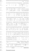
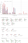
Conger eel has two galectins, termed congerins I and II (Con I and II), that function in mucus as biodefense molecules. Con I and II have acquired a novel protein fold via domain swapping and a new ligand-binding site by accelerated evolution, which enables recognition of some marine bacteria. In this study, we identified a new congerin isotype, congerin P (Con-P), from the peritoneal cells of conger eel. Although Con-P displayed obvious homology with galectins, we observed substitution of 7 out of 8 amino acid residues in the carbohydrate recognition domain that are conserved in all other known galectins. To understand the structure-function relationships of this unique galectin, recombinant Con-P was successfully expressed in Escherichia coli by using a Con II-tagged fusion protein system and subsequently characterized. In the presence of D-mannose, Con-P displayed 30-fold greater hemagglutinating activity than Con I; however, no activity was observed without mannose, indicating that D-mannoside can act as a modulator of Con-P. Frontal affinity chromatography analysis showed that activated Con-P, allosterically induced by mannose, displayed affinity for oligomannose-type sugars as well as N-acetyllactosamine-type β-galactosides. Thus, Con-P represents a new member of the galectin family with unique properties.
Figures


Similar articles
-
Reconstruction of a probable ancestral form of conger eel galectins revealed their rapid adaptive evolution process for specific carbohydrate recognition.Mol Biol Evol. 2007 Nov;24(11):2504-14. doi: 10.1093/molbev/msm185. Epub 2007 Sep 6. Mol Biol Evol. 2007. PMID: 17827170
-
Functional and structural characterization of multiple galectins from the skin mucus of conger eel, Conger myriaster.Comp Biochem Physiol B Biochem Mol Biol. 1999 May;123(1):33-45. doi: 10.1016/s0305-0491(99)00037-1. Comp Biochem Physiol B Biochem Mol Biol. 1999. PMID: 10425711
-
The speciation of conger eel galectins by rapid adaptive evolution.Glycoconj J. 2002;19(7-9):451-8. doi: 10.1023/B:GLYC.0000014074.38755.1d. Glycoconj J. 2002. PMID: 14758068 Review.
-
High-resolution structure of the conger eel galectin, congerin I, in lactose-liganded and ligand-free forms: emergence of a new structure class by accelerated evolution.Structure. 1999 Oct 15;7(10):1223-33. doi: 10.1016/s0969-2126(00)80056-8. Structure. 1999. PMID: 10545323
-
Structure based studies of the adaptive diversification process of congerins.Mol Divers. 2006 Nov;10(4):567-73. doi: 10.1007/s11030-006-9030-8. Mol Divers. 2006. PMID: 16972013 Review.
Cited by
-
Galectin-related protein: An integral member of the network of chicken galectins 1. From strong sequence conservation of the gene confined to vertebrates to biochemical characteristics of the chicken protein and its crystal structure.Biochim Biophys Acta. 2016 Oct;1860(10):2285-97. doi: 10.1016/j.bbagen.2016.06.001. Epub 2016 Jun 3. Biochim Biophys Acta. 2016. PMID: 27268118 Free PMC article.
-
The Lectin Frontier Database (LfDB), and data generation based on frontal affinity chromatography.Molecules. 2015 Jan 8;20(1):951-73. doi: 10.3390/molecules20010951. Molecules. 2015. PMID: 25580689 Free PMC article. Review.
-
Induction of Apoptosis and Antitumor Activity of Eel Skin Mucus, Containing Lactose-Binding Molecules, on Human Leukemic K562 Cells.Mar Drugs. 2015 Jun 19;13(6):3936-49. doi: 10.3390/md13063936. Mar Drugs. 2015. PMID: 26090845 Free PMC article.
-
The zebrafish galectins Drgal1-L2 and Drgal3-L1 bind in vitro to the infectious hematopoietic necrosis virus (IHNV) glycoprotein and reduce viral adhesion to fish epithelial cells.Dev Comp Immunol. 2016 Feb;55:241-252. doi: 10.1016/j.dci.2015.09.007. Epub 2015 Sep 30. Dev Comp Immunol. 2016. PMID: 26429411 Free PMC article.
-
Inhibition of galectins in cancer: Biological challenges for their clinical application.Front Immunol. 2023 Jan 10;13:1104625. doi: 10.3389/fimmu.2022.1104625. eCollection 2022. Front Immunol. 2023. PMID: 36703969 Free PMC article. Review.
References
-
- Kasai K., Hirabayashi J. (1996) Galectins. A family of animal lectins that decipher glycocodes. J. Biochem. 119, 1–8 - PubMed
-
- Perillo N. L., Marcus M. E., Baum L. G. (1998) Galectins. Versatile modulators of cell adhesion, cell proliferation, and cell death. J. Mol. Med. 76, 402–412 - PubMed
-
- Nakamura O., Matsuoka H., Ogawa T., Muramoto K., Kamiya H., Watanabe T. (2006) Opsonic effect of congerin, a mucosal galectin of the Japanese conger, Conger myriaster (Brevoort). Fish Shellfish Immunol. 20, 433–435 - PubMed
-
- Ogawa T., Ishii C., Kagawa D., Muramoto K., Kamiya H. (1999) Accelerated evolution in the protein-coding region of galectin cDNAs, congerin I and congerin II, from skin mucus of conger eel (Conger myriaster). Biosci. Biotechnol. Biochem. 63, 1203–1208 - PubMed
-
- Muramoto K., Kamiya H. (1992) The amino-acid sequence of a lectin from conger eel, Conger myriaster, skin mucus. Biochim. Biophys. Acta 1116, 129–136 - PubMed
MeSH terms
Substances
Associated data
- Actions
LinkOut - more resources
Full Text Sources

