. 2012 Aug;4(8):761-70.
doi: 10.1039/c2mt20086d. Epub 2012 Jul 19.

Changing iron content of the mouse brain during development

Affiliations

Changing iron content of the mouse brain during development

Gregory P Holmes-Hampton et al. Metallomics. 2012 Aug.

Abstract

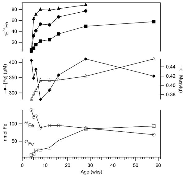
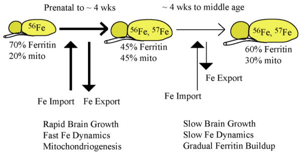
Iron is crucial to many processes in the brain yet the percentages of the major iron-containing species contained therein, and how these percentages change during development, have not been reliably determined. To do this, C57BL/6 mice were enriched in (57)Fe and their brains were examined by Mössbauer, EPR, and electronic absorption spectroscopy; Fe concentrations were evaluated using ICP-MS. Excluding the contribution of residual blood hemoglobin, the three major categories of brain Fe included ferritin (an iron storage protein), mitochondrial iron (consisting primarily of Fe/S clusters and hemes), and mononuclear nonheme high-spin (NHHS) Fe(II) and Fe(III) species. Brains from prenatal and one-week old mice were dominated by ferritin and were deficient in mitochondrial Fe. During the next few weeks of life, the brain grew and experienced a burst of mitochondriogenesis. Overall brain Fe concentration and the concentration of ferritin declined during this burst phase, suggesting that the rate of Fe incorporation was insufficient to accommodate these changes. The slow rate of Fe import and export to/from the brain, relative to other organs, was verified by an isotopic labeling study. Iron levels and ferritin stores replenished in young adult mice. NHHS Fe(II) species were observed in substantial levels in brains of several ages. A stable free-radical species that increased with age was observed by EPR spectroscopy. Brains from mice raised on an Fe-deficient diet showed depleted ferritin iron but normal mitochondrial iron levels.


Figures

Figure 1
Figure 1. Enrichment of 57Fe into 56Fe-enriched mice
Upper plots refer to the % 57Fe scale, including solid triangles (duodenum), circles (average of liver, kidney, heart and spleen) and squares (brain). Solid diamonds in the middle plots refer to total [Fe] in the brain; open triangles refer to brain mass. In the lower plots, open circles and squares refer to nmoles of 56Fe and 57Fe in the brain, respectively. Individual values and statistics are in Tables S1–S3.
Figure 2
Figure 2. Mössbauer spectra of brains isolated at different ages
Spectra were collected at 6 K and with a 0.05 T field applied parallel to the radiation unless otherwise noted. A, 3 wk brain; B, same as A but at 70 K; C, same as A but at 4.3 K and 6 T field applied perpendicular to the radiation; D, 3wk Fe-deficient brain; E, −1wk brain; F, 4 wk brain; G, 58 wk brain. Red lines are composite simulations for EG. Simulations assumed parameters mentioned in the text and percentages in Table 1. The vertical dashed line shows the position of the high-energy line of the central doublet. The arrow in E shows the position of the NHHS FeII feature. Hint values associated with the ferritin sextet ranged from 480 – 506 kG, perhaps reflecting subtle differences in the ferritin core structure.
Figure 3
Figure 3. UV-vis spectra of brains isolated at different ages
A, −1 wk; B, 1 wk; C, 2 wks; D, 3 wks; E, 3 wks and Fe-deficient; F, 4 wks; G, 24 wks; and H, 58 wks. Intensities at wavelengths > 500 nm were multiplied by 5. Contributions due to hemoglobin have not been removed in the displayed spectra.
Figure 4
Figure 4. EPR spectrum of 4 wk brains
Highlighted EPR signals in the bottom panel are indicated by arrows. Conditions: temperature, 4 K; microwave frequency, 9.43 GHz; microwave power, 0.2 mW; modulation amplitude, 10 G; sweep time, 335 sec; time constant, 164 msec. The average of 10 scans is shown. Spectra of brains isolated from animals at other ages are given in Fig. S4. The inset shows the relative intensity of highlighted features as a function of age. Plots are offset by arbitrary amounts.
Figure 5
Figure 5. Model of Fe utilization in the developing mouse brain
During the first ~ 4 wks of life, the mouse brain grows rapidly and exhibits a rapid dynamic exchange of Fe with the blood, including both import and export of Fe to/from the brain. Early during this period most of the Fe in the brain is present as ferritin or ferritin-like material. During the first week or so of life, brain growth and mitochondriogenesis are so fast that Fe cannot be imported rapidly enough to maintain the overall [Fe] in the brain. The decline in [Fe] within neuronal cells causes some ferritin-like Fe to be released, with much of that Fe used for mitochondriogenesis. Gradually, the [Fe] in the brain recovers, rates of dynamic Fe import/export slow, and a greater proportion of Fe becomes stored as ferritin.

Similar articles

Cited by

References

    1. Greenamyre JT, Horowitz MP. Mitochondrial Iron Metabolism and Its Role in Neurodegeneration. J Alzheimers Dis. 2010;20:S551–S568. - PMC - PubMed
    1. Fitzpatrick PF. Tetrahydropterin-dependent amino acid hydroxylases. Annu Rev Biochem. 1999;68:355–381. - PubMed
    1. Ortiz E, Pasquini JM, Thompson K, Felt B, Butkus G, Beard J, Connor JR. Effect of manipulation of iron storage, transport, or availability on myelin composition and brain iron content in three different animal models. J Neurosci Res. 2004;77:681–689. - PubMed
    1. Ramakrishnan U, Rivera J, Villalpando S, Gonzalez-Cossio T, Martorell R, Neufeld LM. Prevalence of anemia and iron deficiency during pregnancy of women supplemented with iron or iron and multiple micronutrients. Faseb Journal. 2001;15:A641–A641.
    1. Beard J, Erikson KM, Jones BC. Neonatal iron deficiency results in irreversible changes in dopamine function in rats. Journal of Nutrition. 2003;133:1174–1179. - PubMed

Publication types