

doi: 10.1038/nchembio.1025.
A new era of GPCR structural and chemical biology
Affiliations
- PMID: 22810761
- PMCID: PMC4031315
- DOI: 10.1038/nchembio.1025
Item in Clipboard
A new era of GPCR structural and chemical biology
Nat Chem Biol.
.
Abstract
G protein-coupled receptors (GPCRs) are versatile molecular machines that regulate the majority of physiological responses to chemically diverse hormones and neurotransmitters. Recent breakthroughs in structural studies have advanced our understanding of GPCR signaling, particularly the selectivity of ligand recognition and receptor activation of G proteins.
Figures
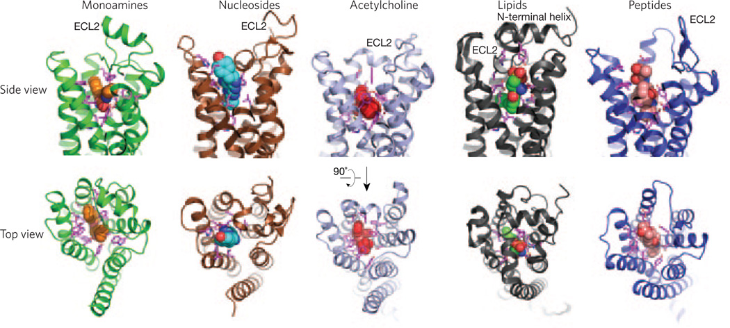
Diversity of ligand binding domains in class A GPCRs. View from the side (top) and from the extracellular surface (bottom) of receptors for chemically diverse ligands. Shown from left to right are the β2-adrenergic receptor bound to carazolol (orange spheres; Protein Data Bank (PDB) code 2RH1), the A2A adenosine receptor bound to ZM24138 (blue spheres; PDB code 3EML), the M2 muscarinic receptor bound to QNB (red spheres; PDB code 3UON), the sphingosine 1-phosphate receptor bound to ML056 (green spheres; PDB code 3V2Y) and the μ-opioid receptor bound to β-FNA (salmon spheres; PDB code 4DKL). A great diversity in extracellular domains, and most notably in ECL2, can be observed. Side chains of residues within 4 Å of the ligands forming the binding pocket are shown in pink.
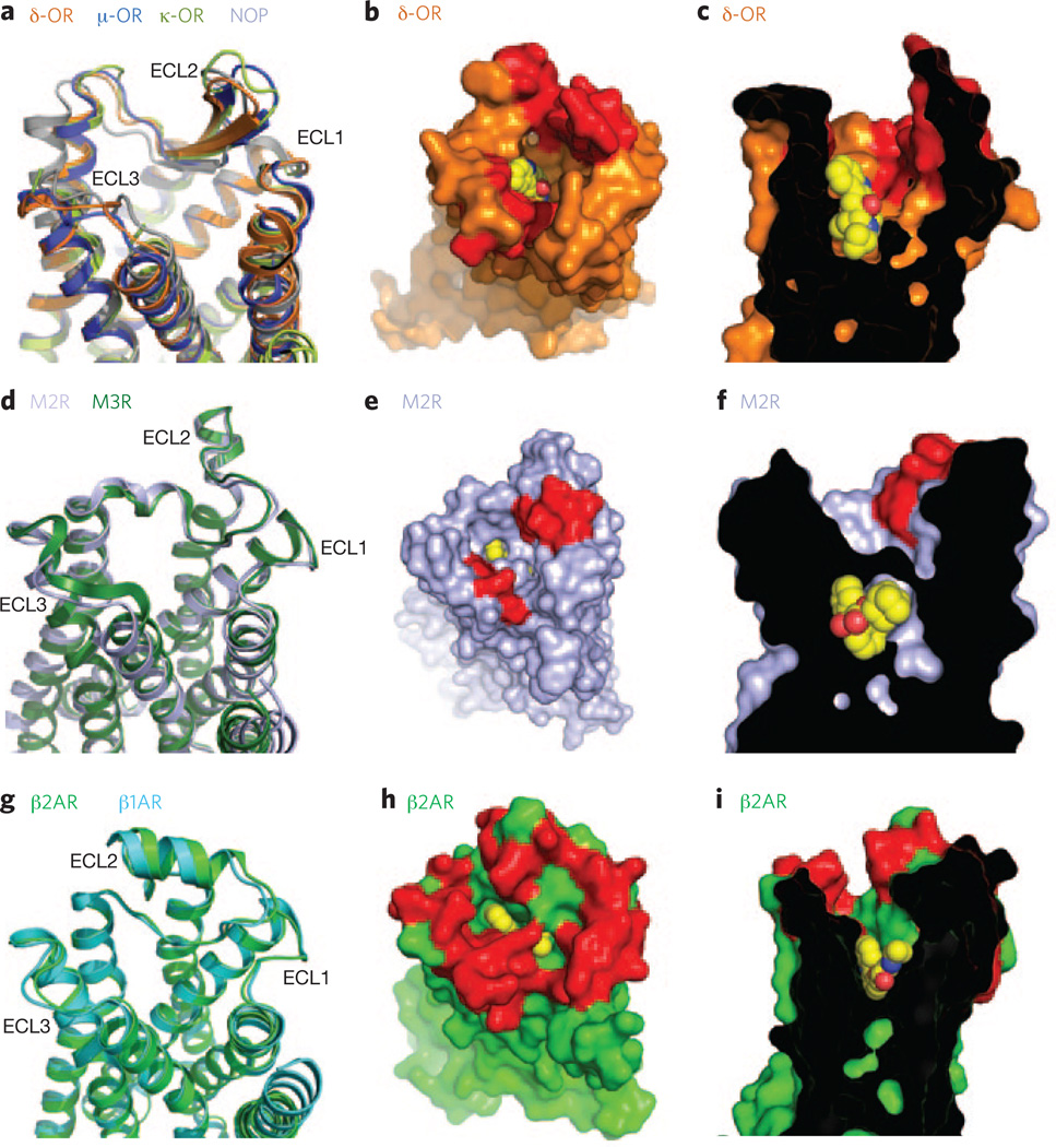
Comparison of binding pockets within receptor subtypes. (a) Extracellular view of the opioid receptors (δ-OR (PDB code 4EJ4), δ-OR (PDB code 4DKL), K-OR (PDB code 4DJH) and nociceptim/ orphanin FQ peptide (NOP) (PDB code 4EA3)) showing structural conservation of the extracellular loops (ECLs 1–3). (b,c) Top (b) and side (c) views of the δ-OR bound to naltrindole (yellow spheres), showing the selectivity determinants in red. (d) Extracellular view of the M2 and M3 muscarinic receptors (M2R (PDB code 3UON) and M3R (PDB code 4DAJ)) showing a similar fold in ECLs 1, 2 and 3. (e,f) Top (e) and side (f) views of the M2R bound to QNB (yellow spheres), with the residues that form the allosteric binding pocket shown in red. (g) Extracellular view of the β1-AR and β2-AR (PDB codes 2Y04 and 2RH1, respectively) showing the structural conservation of extracellular loops (ECLs 1–3). (h,i) Top (h) and side (i) views of the β2-AR bound to carazolol (yellow spheres), with the residues that differ between β1-AR and β2-AR shown in red.
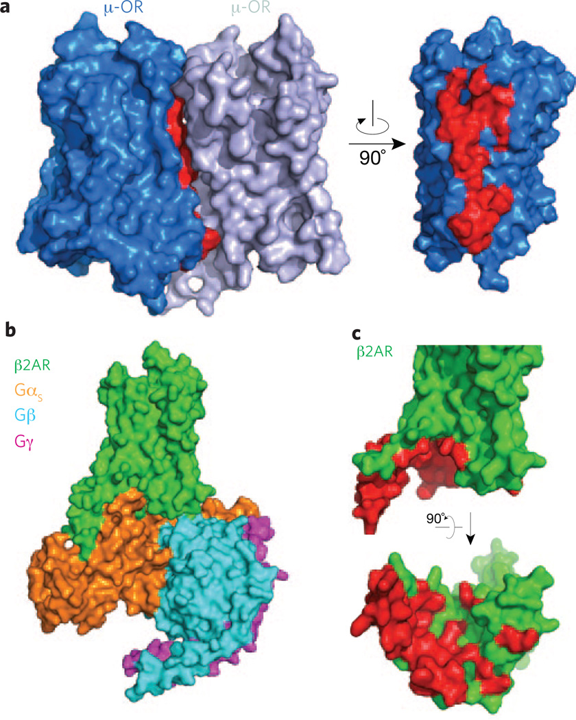
Potential domains to target for regulating receptor function. (a) Dimer interface observed in the δOR crystal structure (PDB code 4DKL) involving transmembrane segments 5 and 6 of each receptor molecule. Residues at the interface are shown in red. (b) Overall view from within the membrane plane of the β2-AR signaling complex (PDB code 3SN6) comprising the β2-AR and the Gs heterotrimer with the αs, β and γ subunits. (c) Side (top) and bottom (bottom) views of β2-AR, with the residues forming the binding pocket of the Gs protein highlighted in red.
References
MeSH terms
Substances
Grants and funding
LinkOut - more resources
Full Text Sources
Other Literature Sources
