

The human fetal lung xenograft: validation as model of microvascular remodeling in the postglandular lung
- PMID: 22811288
- PMCID: PMC3504188
- DOI: 10.1002/ppul.22617
The human fetal lung xenograft: validation as model of microvascular remodeling in the postglandular lung
Abstract
Background: Coordinated remodeling of epithelium and vasculature is essential for normal postglandular lung development. The value of the human-to-rodent lung xenograft as model of fetal microvascular development remains poorly defined.
Aim: The aim of this study was to determine the fate of the endogenous (human-derived) microvasculature in fetal lung xenografts.
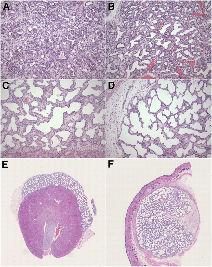
Methods: Lung tissues were obtained from spontaneous pregnancy losses (14-22 weeks' gestation) and implanted in the renal subcapsular or dorsal subcutaneous space of SCID-beige mice (T, B, and NK-cell-deficient) and/or nude rats (T-cell-deficient). Informed parental consent was obtained. Lung morphogenesis, microvascular angiogenesis, and epithelial differentiation were assessed at 2 and 4 weeks post-transplantation by light microscopy, immunohistochemical, and gene expression studies. Archival age-matched postmortem lungs served as control.
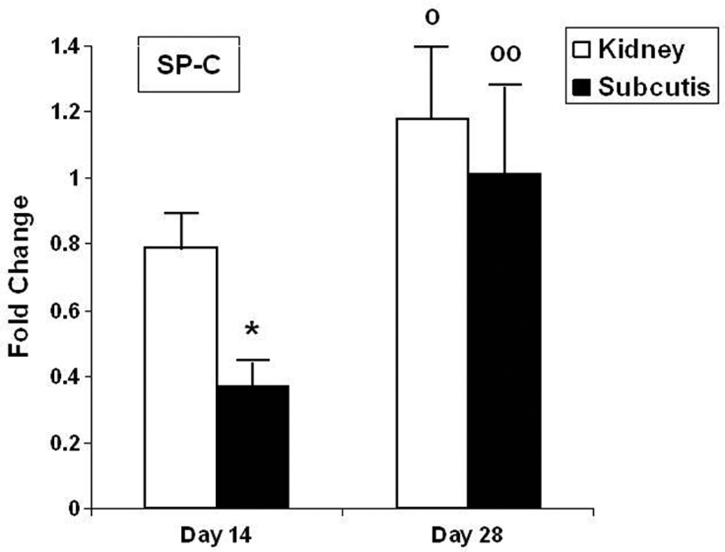
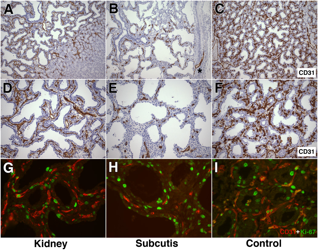
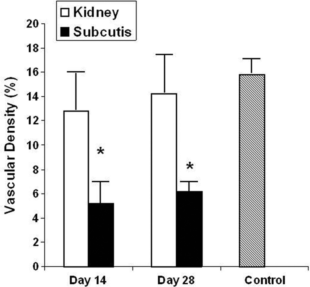
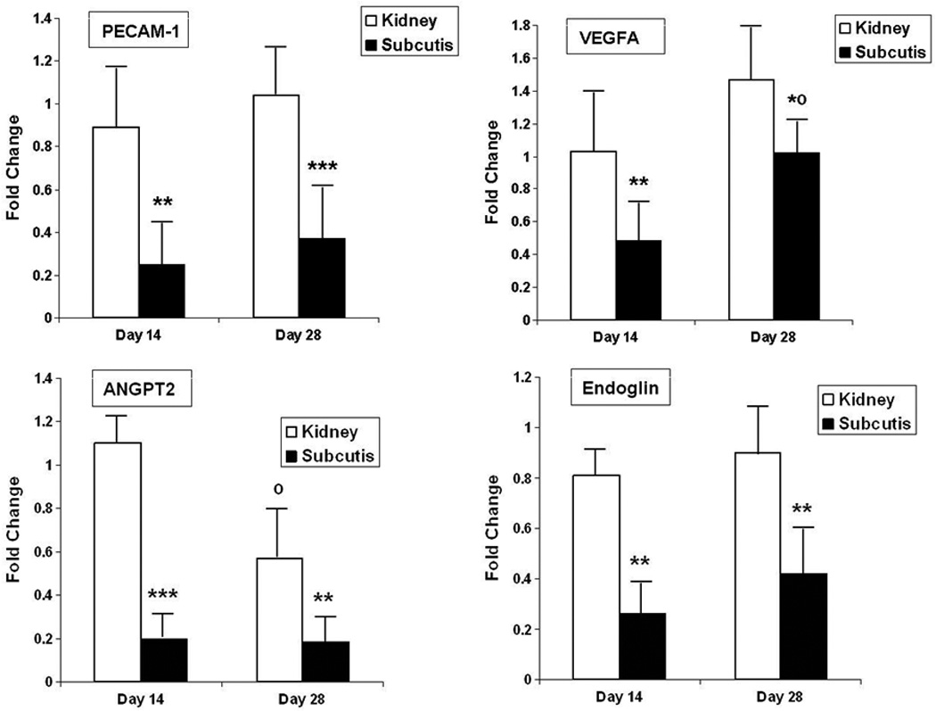
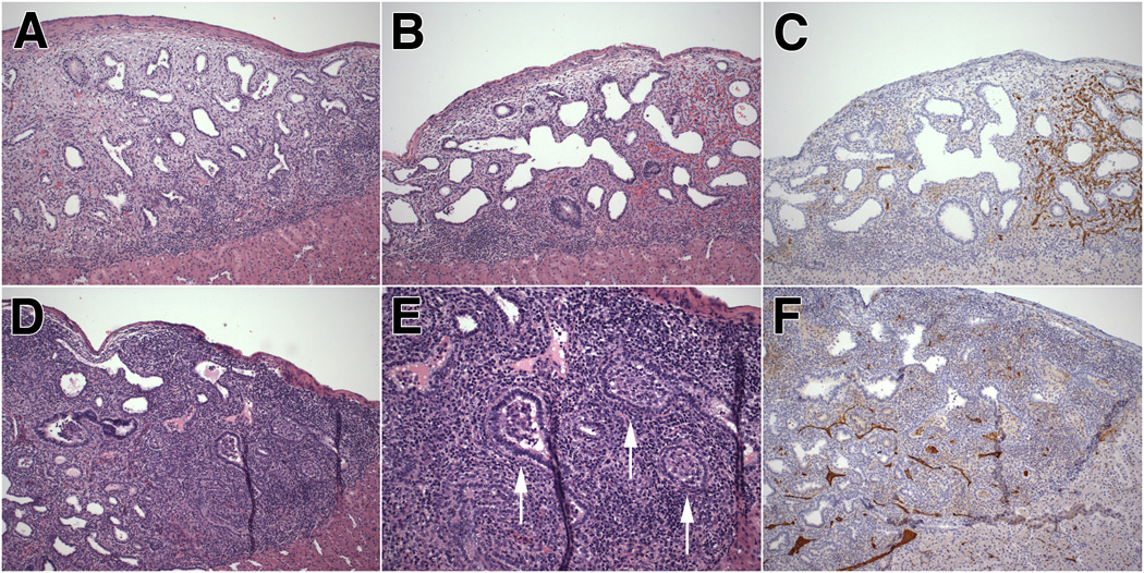
Results: The vascular morphology, density, and proliferation of renal subcapsular grafts in SCID-beige mice were similar to age-matched control lungs, with preservation of the physiologic association between epithelium and vasculature. The microvasculature of subcutaneous grafts in SCID-beige mice was underdeveloped and dysmorphic, associated with significantly lower VEGF, endoglin, and angiopoietin-2 mRNA expression than renal grafts. Grafts at both sites displayed mild airspace dysplasia. Renal subcapsular grafts in nude rats showed frequent infiltration by host lymphocytes and obliterating bronchiolitis-like changes, associated with markedly decreased endogenous angiogenesis.
Conclusion: This study demonstrates the critical importance of host and site selection to ensure optimal xenograft development. When transplanted to severely immune suppressed, NK-cell-deficient hosts and engrafted in the renal subcapsular site, the human-to-rodent fetal lung xenograft provides a valid model of postglandular microvascular lung remodeling.
Copyright © 2012 Wiley Periodicals, Inc.
Conflict of interest statement
There are no conflicts of interest for any of the authors.
Figures
Similar articles
-
Intussusceptive-like angiogenesis in human fetal lung xenografts: Link with bronchopulmonary dysplasia-associated microvascular dysangiogenesis?Exp Lung Res. 2015;41(9):477-88. doi: 10.3109/01902148.2015.1080321. Exp Lung Res. 2015. PMID: 26495956 Free PMC article.
-
Persistent vascular defects in lung allografts attributed to defective endogenous endothelial progenitors.J Surg Res. 2005 Mar;124(1):14-22. doi: 10.1016/j.jss.2004.09.022. J Surg Res. 2005. PMID: 15734474
-
Resilience of the human fetal lung following stillbirth: potential relevance for pulmonary regenerative medicine.Exp Lung Res. 2012 Feb;38(1):43-54. doi: 10.3109/01902148.2011.641139. Epub 2011 Dec 14. Exp Lung Res. 2012. PMID: 22168578 Free PMC article.
-
Human airway xenograft models of epithelial cell regeneration.Respir Res. 2000;1(3):125-8. doi: 10.1186/rr21. Epub 2000 Oct 12. Respir Res. 2000. PMID: 11667974 Free PMC article. Review.
-
Reconstitution of human airway tissue in the humanized xenograft model.J Cyst Fibros. 2004 Aug;3 Suppl 2:63-5. doi: 10.1016/j.jcf.2004.05.014. J Cyst Fibros. 2004. PMID: 15463929 Review.
Cited by
-
Patterns of gene expression and DNA methylation in human fetal and adult liver.BMC Genomics. 2015 Nov 21;16:981. doi: 10.1186/s12864-015-2066-3. BMC Genomics. 2015. PMID: 26589361 Free PMC article.
-
Human lung development: recent progress and new challenges.Development. 2018 Aug 15;145(16):dev163485. doi: 10.1242/dev.163485. Development. 2018. PMID: 30111617 Free PMC article. Review.
-
Xenotransplantation of human fetal adipose tissue: a model of in vivo adipose tissue expansion and adipogenesis.J Lipid Res. 2014 Dec;55(12):2685-91. doi: 10.1194/jlr.D052787. Epub 2014 Sep 5. J Lipid Res. 2014. PMID: 25193996 Free PMC article.
-
Impaired elastin deposition in Fstl1-/- lung allograft under the renal capsule.PLoS One. 2013 Nov 25;8(11):e81368. doi: 10.1371/journal.pone.0081368. eCollection 2013. PLoS One. 2013. PMID: 24282586 Free PMC article.
-
Xenotransplantation models to study the effects of toxicants on human fetal tissues.Birth Defects Res B Dev Reprod Toxicol. 2014 Dec;101(6):410-22. doi: 10.1002/bdrb.21131. Epub 2014 Dec 4. Birth Defects Res B Dev Reprod Toxicol. 2014. PMID: 25477288 Free PMC article. Review.
References
-
- De Paepe ME. Lung growth and development. In: Churg AM, Myers JL, Tazelaar HD, Wright JL, editors. Thurlbeck's pathology of the lung. New York: Thieme Medical Publishers; 2005.
-
- Abman SH. Bronchopulmonary dysplasia: "a vascular hypothesis". Am J Respir Crit Care Med. 2001;164:1755–1756. - PubMed
Publication types
MeSH terms
Grants and funding
LinkOut - more resources
Full Text Sources
