Review
. 2012 Jul;92(3):967-1003.
doi: 10.1152/physrev.00030.2011.

Adaptive and maladaptive cardiorespiratory responses to continuous and intermittent hypoxia mediated by hypoxia-inducible factors 1 and 2

Affiliations
Review

Adaptive and maladaptive cardiorespiratory responses to continuous and intermittent hypoxia mediated by hypoxia-inducible factors 1 and 2

Nanduri R Prabhakar et al. Physiol Rev. 2012 Jul.

Abstract

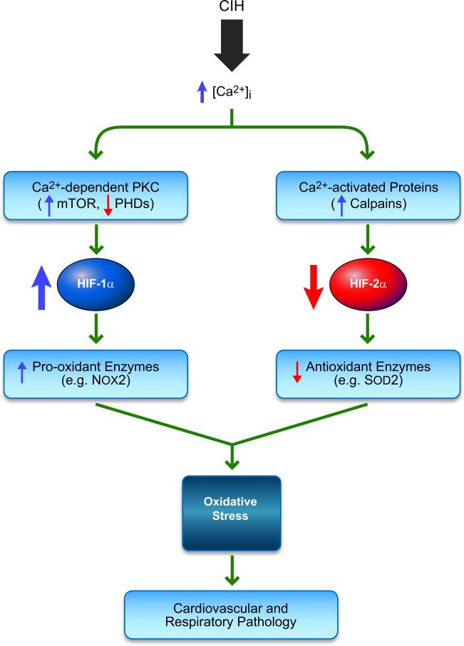
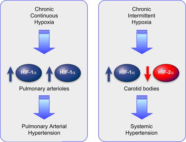
Hypoxia is a fundamental stimulus that impacts cells, tissues, organs, and physiological systems. The discovery of hypoxia-inducible factor-1 (HIF-1) and subsequent identification of other members of the HIF family of transcriptional activators has provided insight into the molecular underpinnings of oxygen homeostasis. This review focuses on the mechanisms of HIF activation and their roles in physiological and pathophysiological responses to hypoxia, with an emphasis on the cardiorespiratory systems. HIFs are heterodimers comprised of an O(2)-regulated HIF-1α or HIF-2α subunit and a constitutively expressed HIF-1β subunit. Induction of HIF activity under conditions of reduced O(2) availability requires stabilization of HIF-1α and HIF-2α due to reduced prolyl hydroxylation, dimerization with HIF-1β, and interaction with coactivators due to decreased asparaginyl hydroxylation. Stimuli other than hypoxia, such as nitric oxide and reactive oxygen species, can also activate HIFs. HIF-1 and HIF-2 are essential for acute O(2) sensing by the carotid body, and their coordinated transcriptional activation is critical for physiological adaptations to chronic hypoxia including erythropoiesis, vascularization, metabolic reprogramming, and ventilatory acclimatization. In contrast, intermittent hypoxia, which occurs in association with sleep-disordered breathing, results in an imbalance between HIF-1α and HIF-2α that causes oxidative stress, leading to cardiorespiratory pathology.


Figures

FIGURE 1
FIGURE 1
Domain structure of hypoxia-inducible factor (HIF) subunits. The following domains are shown: basic helix-loop-helix domain (bHLH), Per-Arnt-Sim homology domain (PAS), O2-dependent degradation domain (ODDD), NH2- and COOH-terminal transactivation domains (TAD-N and TAD-C).
FIGURE 2
FIGURE 2
Multiple HIF-1α isoforms are generated as a result of alternative splicing and alternative promoter utilization. A: alternative splicing of the primary RNA transcript generates mRNAs encoding six different isoforms with the indicated domain composition. B: alternative promoter utilization results in three alternative first exons (Ex 1.1, 1.2, and 1.3) and alternative NH2-terminal amino acid sequences with the translation initiation codon (ATG) present either in the first exon (Ex 1.1, 1.3) or second exon (Ex 1.2).
FIGURE 3
FIGURE 3
Regulation of HIF-1α by O2-dependent hydroxylation. The prolyl hydroxylase PHD2 binds to HIF-1α and catalyzes the hydroxylation of Pro-402 and/or Pro-564. OS-9 stabilizes PHD2-HIF-1α interaction and promotes hydroxylation. VHL binds to prolyl hydroxylated HIF-1α and recruits the ElonginC-ElonginB-CUL2-RBX1 E3-ubiquitin ligase. SSAT2 stabilizes HIF-1α-VHL-ElonginC complex formation, thereby promoting ubiquitination. FIH-1 binds to HIF-1α and catalyzes the hydroxylation of Asn-803, which blocks the binding of the coactivators p300 and CBP to the TAD-C.
FIGURE 4
FIGURE 4
Signal transduction pathways leading to increased HIF-1α protein levels. Activation of protein tyrosine kinases (PTKs) by mutation or ligand binding leads to signaling via the phosphatidylinositol 3-kinase (PI3K) pathway (green) and MAP kinase pathway (purple) that ultimately stimulates the activity of eukaryotic translation initiation factor 4E (eIF-4E) or blocks the activity of its inhibitor, eIF-4E binding protein 1 (4E-BP1), leading to increased translation of HIF-1α mRNA into protein.
FIGURE 5
FIGURE 5
Ubiquitination of HIF-1α by RACK1. Inhibitors of HSP90, such as 17-allylamino-17-demethoxygeldanamycin (17-AAG), block the binding of HSP90 to the PAS-A domain of HIF-1α, allowing RACK1 to bind at this site and recruit the ElonginC ubiquitin ligase complex. Calcineurin dephosphorylates RACK1, thereby preventing its dimerization, which is necessary to bring the ElonginC complex into contact with HIF-1α. Calcineurin activity is inhibited by cyclosporine A, which thereby increases ubiquitination of HIF-1α in a RACK1-dependent manner.
FIGURE 6
FIGURE 6
Ubiquitination of HIF-1α by CHIP. HSP70 recruits CHIP and its associated E2 ligase to HIF-1α.
FIGURE 7
FIGURE 7
Mechanisms for feedback regulation of HIF-1 by target gene products. Stimulatory and inhibitory signals are shown in green and red, respectively. Top panel: in a positive feedback loop, HIF-1 activates the transcription of gene A and other (O) genes. The protein product of gene A stimulates HIF-1 transcriptional activity. Middle panel: in a negative-feedback loop, HIF-1 activates the transcription of gene B. The protein product of gene B inhibits HIF-1 transcriptional activity. Bottom panel: in a mutually antagonistic feedback loop, HIF-1 inhibits the expression of gene C. The protein product of gene C inhibits the activity of HIF-1.
FIGURE 8
FIGURE 8
Regulation of glucose metabolism by HIF-1. The blue boxes indicate products of HIF-1 target genes, which are induced under hypoxic conditions. α-KG, α-ketoglutarate; AcCoA, acetyl coenzyme A; ETC, electron transport chain; TCA, tricarboxylic acid.
FIGURE 9
FIGURE 9
HIF-dependent production of angiogenic growth factors and cytokines in hypoxic cells. Genes encoding the factors shown in purple are directly activated by HIF-1.
FIGURE 10
FIGURE 10
HIF-1-dependent responses to hypoxia in pulmonary artery smooth muscle cells.
FIGURE 11
FIGURE 11
HIF-dependent regulation of iron homeostasis and erthropoiesis.
FIGURE 12
FIGURE 12
Induction of HIF-1 activity by intermittent hypoxia. IH increases the levels of reactive oxygen species (ROS), which activate phospholipase C-γ which converts phosphatidylinositol 4,5-bisphosphate (PIP2) to diacylglycerol (DAG) and inositol 1,4,5-trisphosphate (IP3), resulting in increased intracellular concentration of Ca2+ ([Ca2+]i) and subsequent activation of protein kinase C (PKC) and Ca2+/calmodulin-dependent protein kinase (CaMK). PKC inhibits PHD2 activity, leading to increased HIF-1α protein stability, and stimulates mammalian target of rapamycin (mTOR) activity, leading to increased HIF-1α protein synthesis. CaMK stimulates the binding of p300 to HIF-1α, leading to increased HIF-1-dependent transcriptional activity.
FIGURE 13
FIGURE 13
Differential regulation of HIF-1α and HIF-2α by chronic intermittent hypoxia (CIH) contributes to oxidative stress and cardiorespiratory pathology. CIH increases reactive oxygen species (ROS) via NADPH oxidase-2 (NOX-2) activation. ROS elevate cytosolic calcium [Ca2+]i. Elevated [Ca2+]i leads to increased HIF-1α protein levels as a result of Ca2+-dependent protein kinase C (PKC) activation of mTOR and inhibition of PHDs. Ca2+-activated proteases (calpains) mediate HIF-2α degradation by CIH. HIF-1 activates transcription of the gene encoding the pro-oxidant enzyme NOX-2, whereas decreased HIF-2 activity leads to decreased transcription of genes encoding antioxidant enzymes such as superoxide dismutase 2 (SOD-2). The imbalance between pro-oxidant and anti-oxidant enzymes results in oxidative stress and cardiorespiratory pathology.
FIGURE 14
FIGURE 14
Consequences of responses to chronic continuous hypoxia and chronic intermittent hypoxia in pulmonary arterioles and carotid bodies, respectively.

References

    1. Abud EM, Maylor J, Undem C, Punjabi A, Zaiman AL, Myers AC, Sylvester JT, Semenza GL, Shimoda LA. Digoxin inhibits development of hypoxic pulmonary hyper-tension in mice. Proc Natl Acad Sci USA. 2012;109:1239–1244. - PMC - PubMed
    1. Aprelikova O, Wood M, Tackett S, Chandramouli GV, Barrett JC. Role of ETS transcription factors in the hypoxia-inducible factor-2 target gene selection. Cancer Res. 2006;66:5641–5647. - PubMed
    1. Archer SL, Souil E, Dinh-Xuan AT, Schremmer B, Mercier JC, El Yaagoubi A, Nguyen-Huu L, Reeve HL, Hampl V. Molecular identification of the role of voltage-gated K+ channels, Kv1.5 and Kv21, in hypoxic pulmonary vasoconstriction and control of resting membrane potential in rat pulmonary artery myocytes. J Clin Invest. 1998;101:2319–2330. - PMC - PubMed
    1. Arnardottir ES, Mackiewicz M, Gislason T, Teff KL, Pack AI. Molecular signatures of obstructive sleep apnea in adults: a review and perspective. Sleep. 2009;32:447–470. - PMC - PubMed
    1. Arras M, Ito WD, Scholz D, Winkler B, Schaper J, Schaper W. Monocyte activation in angiogenesis and collateral growth in the rabbit hindlimb. J Clin Invest. 1998;101:40–50. - PMC - PubMed

Publication types

MeSH terms

Substances

LinkOut - more resources