Crystallographic Analysis of Rotavirus NSP2-RNA Complex Reveals Specific Recognition of 5' GG Sequence for RTPase Activity
- PMID: 22811529
- PMCID: PMC3457270
- DOI: 10.1128/JVI.01201-12
Crystallographic Analysis of Rotavirus NSP2-RNA Complex Reveals Specific Recognition of 5' GG Sequence for RTPase Activity
Abstract
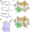
Rotavirus nonstructural protein NSP2, a functional octamer, is critical for the formation of viroplasms, which are exclusive sites for replication and packaging of the segmented double-stranded RNA (dsRNA) rotavirus genome. As a component of replication intermediates, NSP2 is also implicated in various replication-related activities. In addition to sequence-independent single-stranded RNA-binding and helix-destabilizing activities, NSP2 exhibits monomer-associated nucleoside and 5' RNA triphosphatase (NTPase/RTPase) activities that are mediated by a conserved H225 residue within a narrow enzymatic cleft. Lack of a 5' γ-phosphate is a common feature of the negative-strand RNA [(-)RNA] of the packaged dsRNA segments in rotavirus. Strikingly, all (-)RNAs (of group A rotaviruses) have a 5' GG dinucleotide sequence. As the only rotavirus protein with 5' RTPase activity, NSP2 is implicated in the removal of the γ-phosphate from the rotavirus (-)RNA. To understand how NSP2, despite its sequence-independent RNA-binding property, recognizes (-)RNA to hydrolyze the γ-phosphate within the catalytic cleft, we determined a crystal structure of NSP2 in complex with the 5' consensus sequence of minus-strand rotavirus RNA. Our studies show that the 5' GG of the bound oligoribonucleotide interacts extensively with highly conserved residues in the NSP2 enzymatic cleft. Although these residues provide GG-specific interactions, surface plasmon resonance studies suggest that the C-terminal helix and other basic residues outside the enzymatic cleft account for sequence-independent RNA binding of NSP2. A novel observation from our studies, which may have implications in viroplasm formation, is that the C-terminal helix of NSP2 exhibits two distinct conformations and engages in domain-swapping interactions, which result in the formation of NSP2 octamer chains.
Figures


Similar articles
-
Histidine triad-like motif of the rotavirus NSP2 octamer mediates both RTPase and NTPase activities.J Mol Biol. 2006 Sep 22;362(3):539-54. doi: 10.1016/j.jmb.2006.07.050. Epub 2006 Jul 29. J Mol Biol. 2006. PMID: 16934294 Free PMC article.
-
Structure-function analysis of rotavirus NSP2 octamer by using a novel complementation system.J Virol. 2006 Aug;80(16):7984-94. doi: 10.1128/JVI.00172-06. J Virol. 2006. PMID: 16873255 Free PMC article.
-
Crystallographic and biochemical analysis of rotavirus NSP2 with nucleotides reveals a nucleoside diphosphate kinase-like activity.J Virol. 2007 Nov;81(22):12272-84. doi: 10.1128/JVI.00984-07. Epub 2007 Sep 5. J Virol. 2007. PMID: 17804496 Free PMC article.
-
Nonstructural proteins involved in genome packaging and replication of rotaviruses and other members of the Reoviridae.Virus Res. 2004 Apr;101(1):57-66. doi: 10.1016/j.virusres.2003.12.006. Virus Res. 2004. PMID: 15010217 Review.
-
Rotavirus NSP2: A Master Orchestrator of Early Viral Particle Assembly.Viruses. 2024 May 21;16(6):814. doi: 10.3390/v16060814. Viruses. 2024. PMID: 38932107 Free PMC article. Review.
Cited by
-
Molecular characterization of the porcine group A rotavirus NSP2 and NSP5/6 genes from São Paulo State, Brazil, in 2011/12.ScientificWorldJournal. 2013 Jul 15;2013:241686. doi: 10.1155/2013/241686. eCollection 2013. ScientificWorldJournal. 2013. PMID: 23970830 Free PMC article.
-
Structural basis of rotavirus RNA chaperone displacement and RNA annealing.Proc Natl Acad Sci U S A. 2021 Oct 12;118(41):e2100198118. doi: 10.1073/pnas.2100198118. Proc Natl Acad Sci U S A. 2021. PMID: 34615715 Free PMC article.
-
Plasmid-based reverse genetics for probing phosphorylation-dependent viroplasm formation in rotaviruses.Virus Res. 2021 Jan 2;291:198193. doi: 10.1016/j.virusres.2020.198193. Epub 2020 Oct 11. Virus Res. 2021. PMID: 33053412 Free PMC article. Review.
-
Rotavirus viroplasm fusion and perinuclear localization are dynamic processes requiring stabilized microtubules.PLoS One. 2012;7(10):e47947. doi: 10.1371/journal.pone.0047947. Epub 2012 Oct 23. PLoS One. 2012. PMID: 23110139 Free PMC article.
-
Probing the sites of interactions of rotaviral proteins involved in replication.J Virol. 2014 Nov;88(21):12866-81. doi: 10.1128/JVI.02251-14. Epub 2014 Aug 27. J Virol. 2014. PMID: 25165107 Free PMC article.
References
-
- Brunger AT, et al. 1998. Crystallography & NMR system: a new software suite for macromolecular structure determination. Acta Crystallogr. D Biol. Crystallogr. 54:905–921 - PubMed
-
- Chen D, Luongo CL, Nibert ML, Patton JT. 1999. Rotavirus open cores catalyze 5′-capping and methylation of exogenous RNA: evidence that VP3 is a methyltransferase. Virology 265:120–130 - PubMed
Publication types
MeSH terms
Substances
Grants and funding
LinkOut - more resources
Full Text Sources
Research Materials

