. 2012 Oct;86(19):10444-55.
doi: 10.1128/JVI.00986-12. Epub 2012 Jul 18.

Spread of a distinct Stx2-encoding phage prototype among Escherichia coli O104:H4 strains from outbreaks in Germany, Norway, and Georgia

Affiliations

Spread of a distinct Stx2-encoding phage prototype among Escherichia coli O104:H4 strains from outbreaks in Germany, Norway, and Georgia

Lothar Beutin et al. J Virol. 2012 Oct.

Abstract

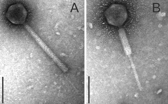
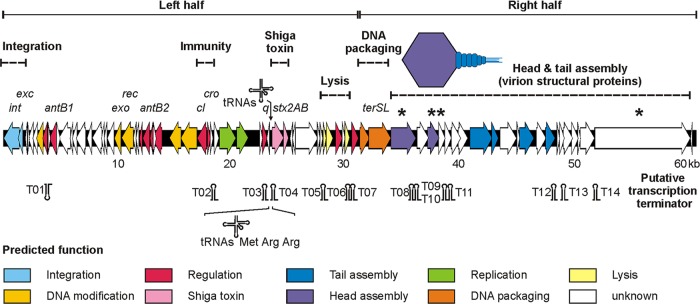
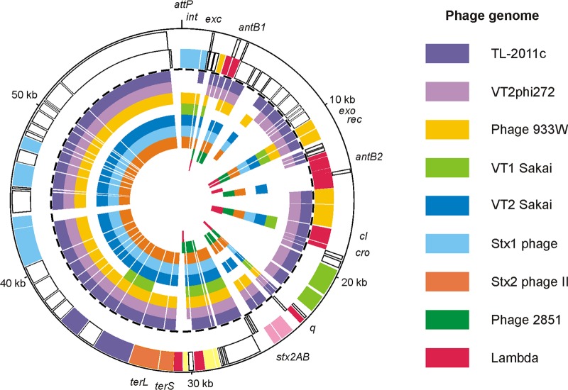
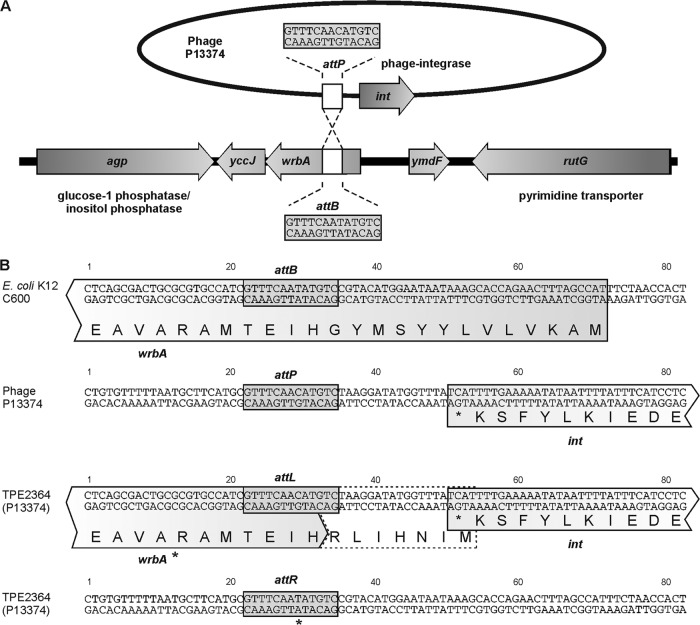
Shiga toxin 2 (Stx2)-producing Escherichia coli (STEC) O104:H4 caused one of the world's largest outbreaks of hemorrhagic colitis and hemolytic uremic syndrome in Germany in 2011. These strains have evolved from enteroaggregative E. coli (EAEC) by the acquisition of the Stx2 genes and have been designated enteroaggregative hemorrhagic E. coli. Nucleotide sequencing has shown that the Stx2 gene is carried by prophages integrated into the chromosome of STEC O104:H4. We studied the properties of Stx2-encoding bacteriophages which are responsible for the emergence of this new type of E. coli pathogen. For this, we analyzed Stx bacteriophages from STEC O104:H4 strains from Germany (in 2001 and 2011), Norway (2006), and the Republic of Georgia (2009). Viable Stx2-encoding bacteriophages could be isolated from all STEC strains except for the Norwegian strain. The Stx2 phages formed lysogens on E. coli K-12 by integration into the wrbA locus, resulting in Stx2 production. The nucleotide sequence of the Stx2 phage P13374 of a German STEC O104:H4 outbreak was determined. From the bioinformatic analyses of the prophage sequence of 60,894 bp, 79 open reading frames were inferred. Interestingly, the Stx2 phages from the German 2001 and 2011 outbreak strains were found to be identical and closely related to the Stx2 phages from the Georgian 2009 isolates. Major proteins of the virion particles were analyzed by mass spectrometry. Stx2 production in STEC O104:H4 strains was inducible by mitomycin C and was compared to Stx2 production of E. coli K-12 lysogens.


Figures

Fig 1
Fig 1
Restriction endonuclease digestion of Stx bacteriophage DNA. Restriction nuclease-digested bacteriophage DNA separated on 0.7% agarose digested with EcoNI (lanes 1 to 5), BsrBI (lanes 6 to 10), and PvuI (lanes 11 to 15). Lanes 1, 6, and 11, phage P8983; 2, 7, and 12, phage P13344; 3, 8, and 13, phage P13374; 4, 9, and 14, phage P13771; and 5, 10, and 15, phage P13772. M, molecular size standard (range, 0.2 to 10 kb).
Fig 2
Fig 2
Stx production of STEC O104:H4 wild-type strains and C600 phage lysogens. Extinction values (OD450) obtained by measuring serial dilutions of the six STEC O104:H4 wild-type strains (Table 1) and five E. coli K-12 lysogens (Table 3) by the Stx EIA. (A) ■, STEC wild-type strains listed in Table 1 grown in the presence of mitomycin C; □, STEC wild-type strains grown without mitomycin C induction. (B) ▲, C600 Stx phage lysogens grown in the presence of mitomycin C; △, C600 Stx phage lysogens grown without mitomycin C.
Fig 3
Fig 3
TEM of Stx phages from STEC O104:H4 strains. Shown are negatively stained phages with a short tail (arrowheads) and hexagonal head isolated from E. coli K-12 (C600) after infection with phage lysates of strains CB8983 (A), CB13344 (B), CB13771 (C), and CB13374 (D). Bars, 50 nm.
Fig 4
Fig 4
TEM of P13374 induction from lysogenic strain TPE2364. Ultrathin sections of two bacterial cells (TPE2364) with maturating virion particles within the cytoplasm indicated by arrows (bars, 500 nm). The cells show signs of bacteriolysis (loss of cytoplasm) (A) and damaged cell walls, as well as the formation of vesicles or opaque clumps (B). (C) TEM of CsCl-purified, negatively stained P13374 particles released by strain TPE2364 (bar, 100 nm). Four phages with a short tail (arrows) and a hexagonal head are shown. In the upper part, two phages with tail-to-tail interactions are shown.
Fig 5
Fig 5
TEM of myovirus phages isolated from the Norwegian STEC O104:H4 strain CB13437. Negatively stained phages comprise a hexagonal head, contractile tail, and kinked tail fibers (Myoviridae). The phages were propagated on E. coli K-12 strain C600 using phage lysates of the Norwegian strain CB13437. (A) Myovirus with noncontracted tail. (B) Myovirus with contracted tail (bars, 100 nm).
Fig 6
Fig 6
Genome organization of prophage P13374. Putative genes are colored according to the predicted functions of their products. The positions of putative Rho-independent transcription terminators and transfer RNAs (tRNAs) are indicated. Further detailed information on the structural components of P13374 are given in Table S3 in the supplemental material. P13374 virion proteins which were identified by SDS-PAGE and mass spectrometry are marked by asterisks.
Fig 7
Fig 7
Relationship of P13374 to Stx phages and phage lambda. The circular synthetic plot shows the P13374 genome organization and the similarity of predicted gene products to proteins of related Stx phages. The two outermost circles represent the plus and minus strands of the P13374 genome, respectively (coloring according to Fig. 6). The inner circles show phage TL-2011c (accession no. JQ011318), phage VT2phi272 (HQ424691), phage 933W (AF125520), phage VT1-Sakai (AP000400), phage VT2-Sakai (AP000422), Stx1-converting phage (AP005153), Stx2-converting phage (AP0044202), phage 2851 (FM180578), and phage lambda (J02459).
Fig 8
Fig 8
Integration of Stx phage P13374 into E. coli K-12 C600. (A) Integration of phage P13374 into the wrbA gene of E. coli C600. Selected genes and the attachment site of the bacteria (attB) and the phage (attP) are indicated. (B) Sequences flanking the attachment sites in E. coli strain C600, phage P13374, and the left (attL) and right (attR) core sequences in lysogenic C600 strains. wrbA* indicates a putative modified wrbA gene.

References

    1. Altschul SF, et al. 1997. Gapped BLAST and PSI-BLAST: a new generation of protein database search programs. Nucleic Acids Res. 25:3389–3402 - PMC - PubMed
    1. Aurass P, Prager R, Flieger A. 2011. EHEC/EAEC O104:H4 strain linked with the 2011 German outbreak of haemolytic uremic syndrome enters into the viable but non-culturable state in response to various stresses and resuscitates upon stress relief. Environ. Microbiol. 13:3139–3148 - PubMed
    1. Baker DR, Moxley RA, Francis DH. 1997. Variation in virulence in the gnotobiotic pig model of O157:H7 Escherichia coli strains of bovine and human origin. Adv. Exp. Med. Biol. 412:53–58 - PubMed
    1. Beutin L, Martin A. 2012. Outbreak of Shiga toxin-producing Escherichia coli (STEC) O104:H4 infection in Germany causes a paradigm shift with regard to human pathogenicity of STEC strains. J. Food Prot. 75:408–418 - PubMed
    1. Beutin L, et al. 2007. Comparative evaluation of the Ridascreen verotoxin enzyme immunoassay for detection of Shiga-toxin producing strains of Escherichia coli (STEC) from food and other sources. J. Appl. Microbiol. 102:630–639 - PubMed

Publication types

MeSH terms

Associated data

LinkOut - more resources