. 2012 Nov 29;87(5):127.
doi: 10.1095/biolreprod.112.100735. Print 2012 Nov.

Transforming growth factor Beta 1 stimulates profibrotic activities of luteal fibroblasts in cows

Affiliations

Transforming growth factor Beta 1 stimulates profibrotic activities of luteal fibroblasts in cows

Dulce Maroni et al. Biol Reprod. .

Abstract

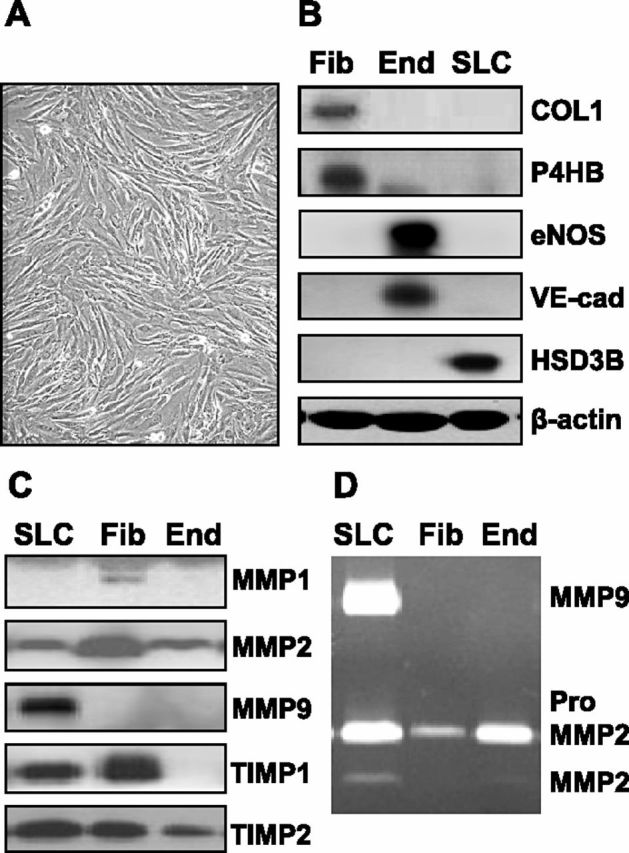
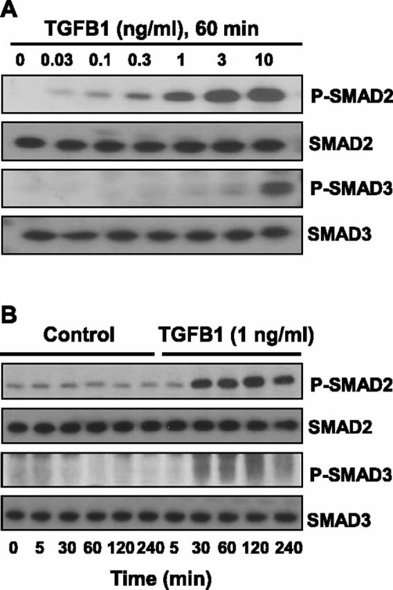
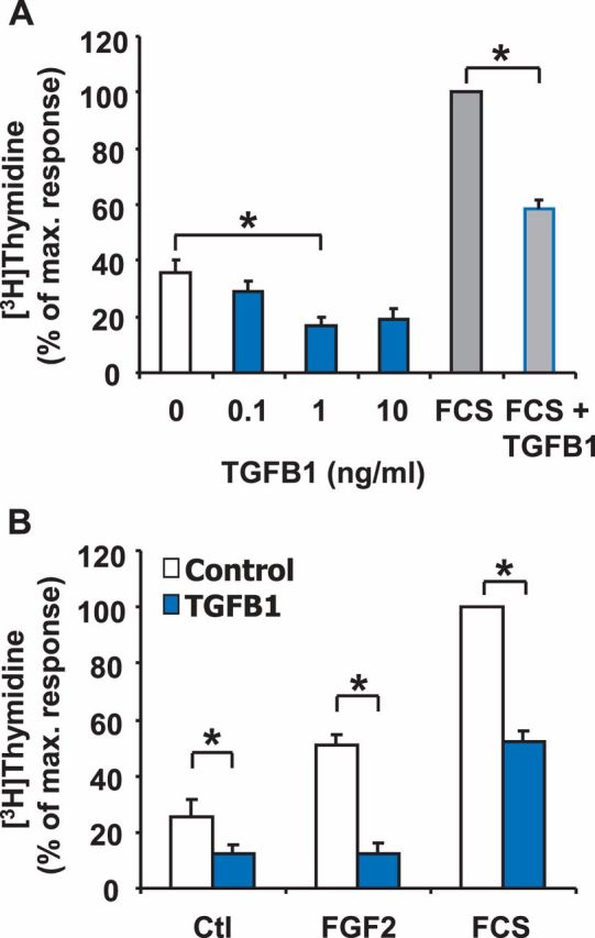
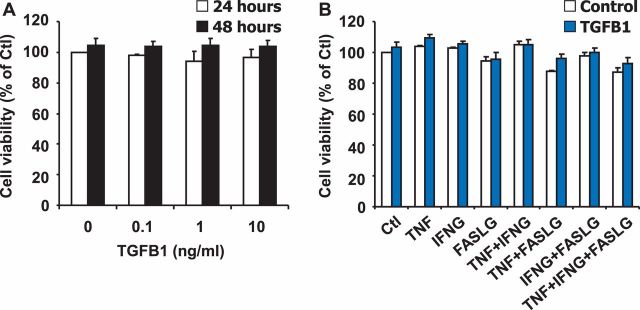
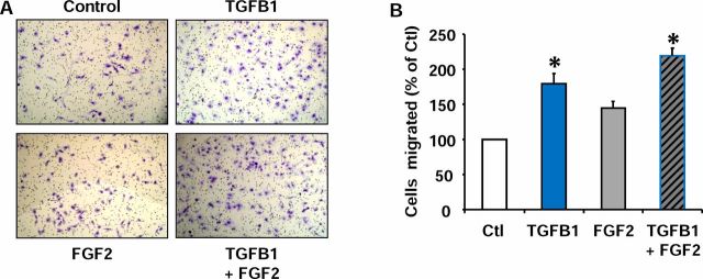
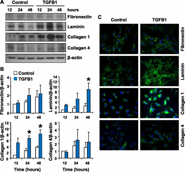
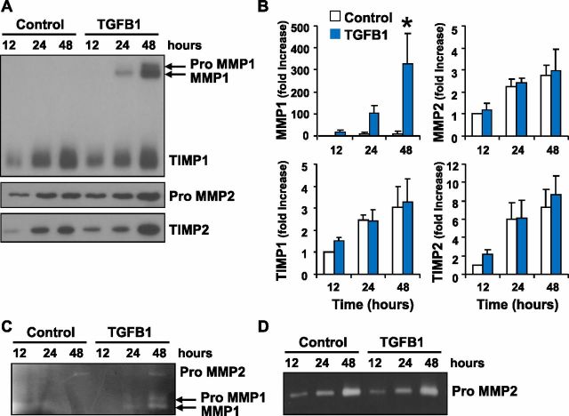
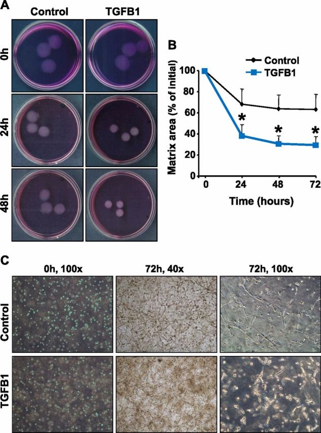
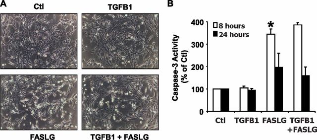
Luteolysis is characterized by angioregression, luteal cell apoptosis, and remodeling of the extracellular matrix characterized by deposition of collagen 1. Transforming growth factor beta 1 (TGFB1) is a potent mediator of wound healing and fibrotic processes through stimulation of the synthesis of extracellular matrix components. We hypothesized that TGFB1 stimulates profibrotic activities of luteal fibroblasts. We examined the actions of TGFB1 on luteal fibroblast proliferation, extracellular matrix production, floating gel contraction, and chemotaxis. Fibroblasts were isolated from the bovine corpus luteum. Western blot analysis showed that luteal fibroblasts expressed collagen 1 and prolyl 4-hydroxylase but did not express markers of endothelial or steroidogenic cells. Treatment of fibroblasts with TGFB1 stimulated the phosphorylation of SMAD2 and SMAD3. [(3)H]thymidine incorporation studies showed that TGFB1 caused concentration-dependent reductions in DNA synthesis in luteal fibroblasts and significantly (P < 0.05) reduced the proliferative effect of FGF2 and fetal calf serum. However, TGFB1 did not reduce the viability of luteal fibroblasts. Treatment of luteal fibroblasts with TGFB1 induced the expression of laminin, collagen 1, and matrix metalloproteinase 1 as determined by Western blot analysis and gelatin zymography of conditioned medium. TGFB1 increased the chemotaxis of luteal fibroblasts toward fibronectin in a transwell system. Furthermore, TGFB1 increased the fibroblast-mediated contraction of floating bovine collagen 1 gels. These results suggest that TGFB1 contributes to the structural regression of the corpus luteum by stimulating luteal fibroblasts to remodel and contract the extracellular matrix.


Figures

Fig. 1
Fig. 1
Characterization of fibroblasts isolated from the bovine corpus luteum. Luteal fibroblasts were isolated from bovine corpus luteum and characterized by their morphology and expression of cell markers. Fibroblasts were cultured in monolayer in growth medium (DMEM supplemented with 10% FCS). Supernatant medium was harvested, and the expression of matrix-remodeling proteins was examined by Western blot analysis and zymography. A) Luteal fibroblasts display typical fibroblast morphology; they are spindle-shaped, with numerous cytoplasmic processes forming a network of intercellular contacts. Original magnification ×100. B) Characterization of luteal fibroblasts by Western blot analysis of cell lysates (40 μg protein/lane). Luteal endothelial cell (End) and steroidogenic luteal cell (SLC) protein extracts were prepared as previously described [30, 37]. Luteal fibroblasts (Fib) expressed fibroblast cell markers collagen 1 (COL1) and prolyl 4-hydroxylase (P4HB) but did not express endothelial cell (End) markers VE-cad and eNOS. In addition, luteal fibroblasts did not express the SLC marker HSD3B. C) The expression of matrix-remodeling proteins by luteal fibroblasts was examined by Western blot analysis of conditioned medium. Luteal fibroblasts secreted MMP1, MMP2, TIMP1, and TIMP2 but did not express MMP9. D) Gelatin zymography of conditioned medium (30 μl/lane) shows the presence of MMP2 zymogen in conditioned medium from cultured luteal fibroblasts.
Fig. 2
Fig. 2
TGFB1 induces phosphorylation of SMAD2 and SMAD3 in luteal fibroblasts. A) Concentration response to TGFB1. Cells were serum-starved and treated with TGFB1 (0–10 ng/ml) for 60 min under serum-free conditions. B) Time-course response to TGFB1. Cells were serum-starved and treated with TGFB1 (1 ng/ml) for up to 4 h under serum-free conditions. Levels of phosphorylated SMAD2 and SMAD3 were determined by Western blot analysis of cell lysates (30 μg protein/lane). Levels of total SMAD2 and SMAD3 proteins are also shown.
Fig. 3
Fig. 3
TGFB1 reduces DNA synthesis in luteal fibroblasts. A) [3H]thymidine incorporation assay of fibroblasts plated at low density and treated with or without TGFB1 (0–10 ng/ml) for 24 h. Some cells were cultured in the presence of 5% FCS alone or in combination with TGFB1 (1 ng/ml) for 24 h. Data are expressed as the percentage incorporation of [3H]thymidine compared to the maximal response group (5% FCS). Data represent three independent experiments, each performed in triplicate (mean ± SEM, n = 3, *P < 0.05). B) [3H]thymidine incorporation assay of fibroblasts plated at low density and treated for 24 h with or without TGFB1 (1 ng/ml) in the absence (Ctl) or presence of FGF2 (10 ng/ml) and 5% FCS. Data are expressed as the percentage incorporation of [3H]thymidine compared to the maximal response group (5% FCS). Data represent three independent experiments, each performed in triplicate (mean ± SEM, n = 3, *P < 0.05).
Fig. 4
Fig. 4
TGFB1 does not reduce viability of luteal fibroblasts. A) MTT assay of fibroblasts plated on plastic at high density and treated for 24 and 48 h without or with TGFB1 (0–10 ng/ml) in the absence of FCS. Data represent three independent experiments, each performed in triplicate (mean ± SEM, n = 3, P > 0.05 vs. 0 ng/ml of TGFB1). B) MTT assay of fibroblasts plated on plastic at high density and treated for 48 h with control medium (Control) or TGFB1 (1 ng/ml), alone or in combination with control medium (Ctl), TNF (50 ng/ml), IFNG (50 ng/ml), or FASLG (100 ng/ml) in the absence of FCS. Results are expressed as the percentage absorbance observed in the control group. Data shown represent three independent experiments, each performed in triplicate (mean ± SEM, n = 3, P > 0.05 vs. Ctl).
Fig. 5
Fig. 5
TGFB1 increases chemotaxis of luteal fibroblasts toward fibronectin. Fibroblasts were harvested with trypsin and used for chemotaxis in the blind-well Boyden chamber assay. Fibroblasts were treated with control medium (Ctl), TGFB1 (1 ng/ml), or FGF2 (10 ng/ml) alone or in combination in the upper well. Fibronectin (20 μg/ml) was used as the chemoattractant. Chemotaxis was measured as the number of migrated cells after a 6-h incubation period. A) Migrated fibroblasts found on the bottom of the transwell membrane were fixed, stained, and photographed. Original magnification ×100. B) Quantification of cell migration. Cell migration is represented as a percentage of the number of fibroblasts migrated in the control treatment group. Data shown represent three independent experiments, each performed in triplicate (mean ± SEM, n = 3, *P < 0.05 vs. Ctl).
Fig. 6
Fig. 6
Effect of TGFB1 treatment on the expression of extracellular matrix proteins by luteal fibroblasts. Luteal fibroblasts were treated with control medium (Control) or TGFB1 (1 ng/ml) in serum-free medium for up to 48 h, and extracellular matrix protein expression was examined by Western blot analysis and immunofluorescence. A) Western blot analysis of cell lysates (40 μg protein/lane). A representative Western blot shows levels of fibronectin, laminin, collagen 1, and collagen 4, with β-actin as loading control. B) Data from densitometric analysis are shown as fold-increases over the untreated control samples after normalization to β-actin. Treatment with TGFB1 up-regulated the expression of laminin and collagen 1. Data represent three separate experiments (mean ± SEM, n = 3. *P < 0.05 vs. Control). C) Analysis of protein expression by immunofluorescence of luteal fibroblast treated with TGFB1 for 24 h. TGFB1 treatment increased the intensity of the staining for laminin and collagen 1. Original magnification ×200.
Fig. 7
Fig. 7
Effect of TGFB1 on MMP release by luteal fibroblasts. Fibroblasts were cultured in monolayer and treated with TGFB1 (1 ng/ml) in serum-free medium. Supernatant medium was harvested, and the expression of matrix-remodeling proteins was examined by Western blot analysis and zymography. A) Western blot analysis (30 μl medium/lane) for MMP1, TIMP1, MMP2, and TIMP2. B) Data from densitometric analysis shown as fold-increases over the untreated control samples (12 h). TGFB1 up-regulated the expression of latent and active MMP1. Data represent three separate experiments (mean ± SEM, n = 3, *P < 0.05 vs. Control). C) Casein zymography (30 μl medium/lane) for MMP1. The presence of the active form of MMP1 was detected in medium samples from TGFB1-treated cells only. D) Gelatin zymography (30 μl medium/lane) for MMP2. Luteal fibroblasts produced only inactive MMP2.
Fig. 8
Fig. 8
TGFB1 enhanced contraction of fibroblast-populated collagen 1 matrices. The role of TGFB1 in luteal fibroblast contractile function was investigated using fibroblast-populated bovine collagen 1 matrices. Fibroblasts were harvested with trypsin and cast into collagen matrices, which were then floated in DMEM with or without TGFB1 (1 ng/ml). A) Representative experiment using control (top) and TGFB1 (1 ng/ml; bottom)-treated collagen matrices. B) Matrix size is expressed as the percentage of initial area. Quantitative analysis demonstrated that matrix area was significantly smaller in matrices treated with TGFB1. Data represent three independent experiments, each performed in triplicate (mean ± SEM, n = 3, *P < 0.05 vs. Control). C) Morphology of fibroblasts cast into floating collagen matrices. Fibroblasts within collagen matrices produce a dendritic network of cytoplasmic extensions. Pictures were taken using a phase-contrast microscope at the indicated culture time points and magnification.
Fig. 9
Fig. 9
Effect of TGFB1 on fibroblasts plated on collagen 1 gels. A) Morphology of fibroblasts plated (5 × 10⁴ cells/well) in growth medium on 48-well plates coated with bovine collagen 1 gels (0.15 ml, ∼2.4 mg/ml). Plated cells were treated with control medium (Ctl), TGFB1 (1 ng/ml), FASLG (100 ng/ml), or their combination. Pictures were taken under a phase-contrast microscope after 24 h of incubation at 37°C. Original magnification ×200. B) Caspase 3 activity was measured after 24 h of treatment using the Caspase-Glo 3/7 assay kit. Data are expressed as a percentage of the caspase activity observed in controls and represent three independent experiments, each performed in triplicate with similar results (mean ± SEM, n = 3, *P < 0.05).

References

    1. Niswender GD, Juengel JL, Silva PJ, Rollyson MK, McIntush EW.. Mechanisms controlling the function and life span of the corpus luteum. Physiol Rev 2000; 80:1–29. - PubMed
    1. Duncan WC, McNeilly AS, Illingworth PJ.. The effect of luteal “rescue” on the expression and localization of matrix metalloproteinases and their tissue inhibitors in the human corpus luteum. J Clin Endocrinol Metab 1998; 83:2470–2478. - PubMed
    1. Smith MF, Ricke WA, Bakke LJ, Dowb MPD, Smith GW.. Ovarian tissue remodeling: role of matrix metalloproteinases and their inhibitors. Mol Cell Endocrinol 2002; 191:45–56. - PubMed
    1. Ribeiro LA, Turba ME, Zannoni A, Bacci ML, Forni M.. Gelatinases, endonuclease and vascular endothelial growth factor during development and regression of swine luteal tissue. BMC Dev Biol 2006; 6:58–66. - PMC - PubMed
    1. Stocco C, Telleria C, Gibori G.. The molecular control of corpus luteum formation, function, and regression. Endocr Rev 2007; 28:117–149. - PubMed

Publication types

MeSH terms