. 2012 Sep 15;18(18):5020-30.
doi: 10.1158/1078-0432.CCR-12-1072. Epub 2012 Jul 18.

Ras-driven transcriptome analysis identifies aurora kinase A as a potential malignant peripheral nerve sheath tumor therapeutic target

Affiliations

Ras-driven transcriptome analysis identifies aurora kinase A as a potential malignant peripheral nerve sheath tumor therapeutic target

Ami V Patel et al. Clin Cancer Res. .

Abstract

Purpose: Patients with neurofibromatosis type 1 (NF1) develop malignant peripheral nerve sheath tumors (MPNST), which are often inoperable and do not respond well to current chemotherapies or radiation. The goal of this study was to use comprehensive gene expression analysis to identify novel therapeutic targets.

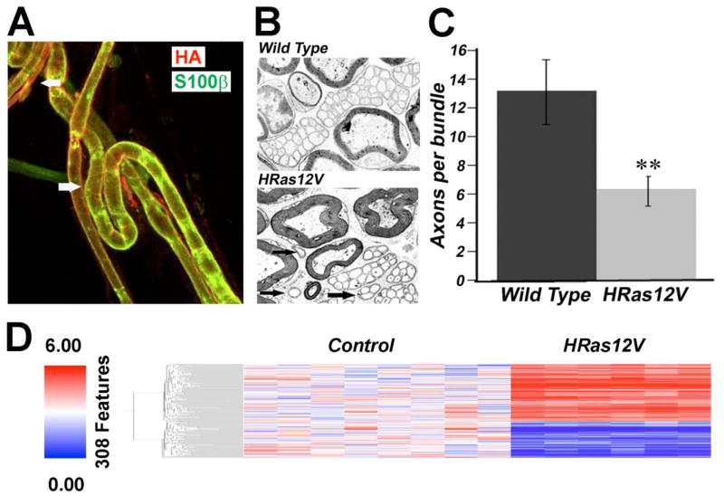
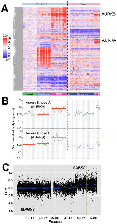
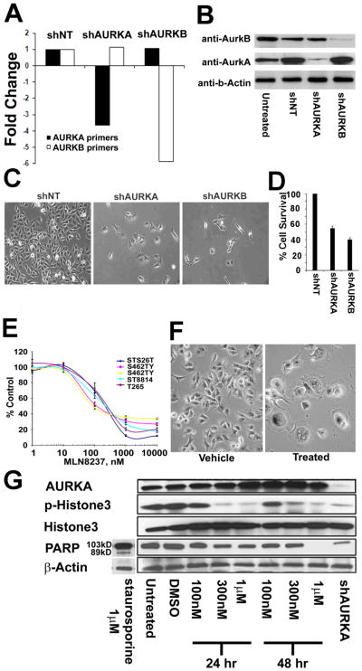
Experimental design: Nerve Schwann cells and/or their precursors are the tumorigenic cell types in MPNST because of the loss of the NF1 gene, which encodes the RasGAP protein neurofibromin. Therefore, we created a transgenic mouse model, CNP-HRas12V, expressing constitutively active HRas in Schwann cells and defined a Ras-induced gene expression signature to drive a Bayesian factor regression model analysis of differentially expressed genes in mouse and human neurofibromas and MPNSTs. We tested functional significance of Aurora kinase overexpression in MPNST in vitro and in vivo using Aurora kinase short hairpin RNAs (shRNA) and compounds that inhibit Aurora kinase.

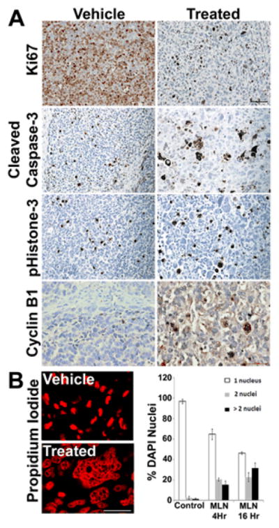
Results: We identified 2,000 genes with probability of linkage to nerve Ras signaling of which 339 were significantly differentially expressed in mouse and human NF1-related tumor samples relative to normal nerves, including Aurora kinase A (AURKA). AURKA was dramatically overexpressed and genomically amplified in MPNSTs but not neurofibromas. Aurora kinase shRNAs and Aurora kinase inhibitors blocked MPNST cell growth in vitro. Furthermore, an AURKA selective inhibitor, MLN8237, stabilized tumor volume and significantly increased survival of mice with MPNST xenografts.

Conclusion: Integrative cross-species transcriptome analyses combined with preclinical testing has provided an effective method for identifying candidates for molecular-targeted therapeutics. Blocking Aurora kinases may be a viable treatment platform for MPNST.


Conflict of interest statement

Conflicts of Interest: None

Figures

Figure 1
Figure 1. Schwann Cell-specific Ras-driven Gene Expression
(A) Immunolabeling adult teased nerve fibers detected HRas (as detected by anti-HA, red) at the membrane of myelinated fibers (bottom: white arrow) and smaller unmyelinated fibers (top: white arrow). Schwann cell cytoplasm is counterstained with anti-S100 (green), which preferentially labels myelinating Schwann cells. (B) Electron micrographs of a cross-section through saphenous nerves from adult wild type or CNP-HRAS12V mice are shown. At high magnification, the unmyelinated axons with accompanying Schwann cell processes in the HRAS12V mutant showed normal ensheathment but few fibers per bundle. Groups of unmyelinated axons in sapphenous nerve contained diminished numbers of axons in the CNP-HRAS12V mutant. (C) At 1 year old, 13.0 +/− 2.2 axons were grouped by a single non-myelin forming Schwann cell in wild type mice, but only 6.4 +/− 0.9 in CNP-V12Ras mutants (P ≤ 0.004). Black arrows point to small groups of unmyelinated axons in mutant. The phenotype did not worsen with age (not shown). (D) Schwann-cell specific Ras signature in CNP-HRAS12V transgenic mouse model.
Figure 2
Figure 2. AURKA is Overexpressed and Amplified in MPNST
(A) Bayesian latent factor regression modeling of microarray gene expression data identified 2,000 gene orthologs linked to the Ras pathway in mouse and human NF1 tumors. (B) AURKA and AURKB were expressed at high levels in MPNST cell lines and primary tumors relative to neurofibromas and normal nerves. Each sample is represented by a vertical line and is directly under the sample type categories represented in (A). Sample types categories are separated by an open space. The vertical dashed line divides mouse samples (left) and human samples (right). Expression level of AURKA (top) or AURKB (bottom) was normalized to mouse or human normal nerve, respectively, and represented as 1.00 on a log scale. The red line represents the average expression level among samples within each sample type (C) Example of SNP-array analysis of an MPNST. This figure shows the log R ratio (LRR) of chromosome 20 for tumor MPNST K (see Supplementary Table S1). A blue line denotes the running average of LRR values. A value of 0 indicates the presence of two copies of each locus analyzed. A mean value above 0 indicates a copy number gain of the locus interrogated. A vertical yellow line localizes the AURKA locus.
Figure 3
Figure 3. Blocking AURK gene expression reduces MPNST cell survival in vitro MLN8237 reduces AURKA activity and MPNST cell survival in vitro
(A) AURKA expression was diminished by AURKA shRNA; AURKB expression was diminished by AURKB shRNA. Blocking each AURK expression at the RNA level was specific as determined by qRT-PCR. (B) Blocking expression of either AURK reduced protein levels of both as detected by Western blot. (C) Phase contrast images of MPNST cells showed a dramatic reduction in cell number after treating with shAURKA or shAURKB. (D) Quantification of cell accumulation by MTS assay showed decreased survival in response to shAURKA and shAURKB. (E) MPNST cells became senescent 48hrs post treatment with MLN8237 as compared to vehicle treated cells as seen in these phase contract images. (F) MLN8237 Aurora kinase A inhibitor reduced survival of all 5 MPNST cell lines tested at an average IC50 of 164nm. (G) Protein levels for AurkA, p-Histone3, total Histone 3, PARP and loading control β-actin for MPNST cells (S462TY) treated with 100nM, 300nM and 1μM of MLN8237 collected at 24hr and 48hr post treatment with drug. Untreated and vehicle treated were used as a negative control and shAURKA treated MPNST cells collected at 4 days post virus treatment serve as a positive control.
Figure 4
Figure 4. MLN8237 reduces Tumor Volume and Increases Survival of Mice with MPNST Xenografts
(A – C) Inhibiting Aurora kinase A diminished tumor volume. (D) Kaplan-Meier curve shows increased survival of mice with MPNST xenografts in response to MLN8237 (p=0.0005); median survival for vehicle-treated mice was 41 days; median survival for MLN8237-treated mice was undefined. (E) Pharmacokinetic analysis of MLN8237 in MPNST xenografts. Average blood plasma concentrations of MLN8237 (nM) in 3 control nude mice at 2, 4, and 16 hours after dosing are plotted on a log scale. Dashed line represents dose (1μM) required for Aurora kinase inhibition in vivo.
Figure 5
Figure 5. MLN8237 induces cytomegaly in MPNST Xenografts
(A) MPNST xenografts treated with vehicle control or MLN8237 and assayed for Ki67, a marker of cell proliferation, p-Histone 3, a marker of proliferation, cleaved caspase 3, a marker for apoptosis, or cyclinB1, a marker of cell in the G2/M phase of the cell cycle. Vehicle and MLN8237-treated mice showed similar levels of p-histone 3 and cleaved caspase 3. Noticeably fewer cells were positively labeled for Ki67 in the MLN-8237 treated tissue samples as compared to Vehicle. There was a marked increase in the positive staining for CyclinB1 in the MLN8237 treated tissue samples compared to vehicle treated samples. (B) Many multinucleated cells were detected in MLN8237-treated xenografts, as visualized with propidium iodide and quantified by counting DAPI-stained nuclei per cell at 4 hours and 16 hours.

Similar articles

Cited by

References

    1. Katz D, Lazar A, Lev D. Malignant peripheral nerve sheath tumour (MPNST): the clinical implications of cellular signalling pathways. Expert Rev Mol Med. 2009;11:e30. - PubMed
    1. Bottillo I, Ahlquist T, Brekke H, Danielsen SA, van den Berg E, Mertens F, et al. Germline and somatic NF1 mutations in sporadic and NF1-associated malignant peripheral nerve sheath tumours. J Pathol. 2009;217:693–701. - PubMed
    1. Sherman LS, Atit R, Rosenbaum T, Cox AD, Ratner N. Single cell Ras-GTP analysis reveals altered Ras activity in a subpopulation of neurofibroma Schwann cells but not fibroblasts. Journal of Biological Chemistry. 2000;275:30740–30745. - PMC - PubMed
    1. Basu TN, Gutmann DH, Fletcher JA, Glover TW, Collins FS, Downward J. Aberrant regulation of ras proteins in malignant tumour cells from type 1 neurofibromatosis patients. Nature. 1992;356:663–664. - PubMed
    1. DeClue JE, Papageorge AG, Fletcher JA, Diehl SR, Ratner N, Vass WC, et al. Abnormal regulation of mammalian p21ras contributes to malignant tumor growth in von Recklinghausen (Type 1) neurofibromatosis. Cell. 1992;69:265–273. - PubMed

MeSH terms