. 2012 Mar;3(1):28-40.
doi: 10.3978/j.issn.2078-6891.2012.006.

Techniques of hepatic resection

Affiliations

Techniques of hepatic resection

Robert J Aragon et al. J Gastrointest Oncol. 2012 Mar.

Abstract

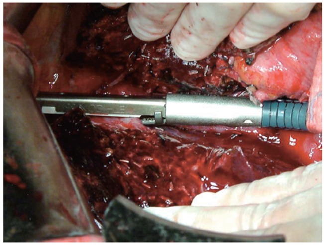
Liver resections are high risk procedures performed by experienced surgeons. The role of liver resection in malignant disease has changed over the last 100 years with great improvement in morbidity, mortality and long term survival. New understanding in liver anatomy, improved perioperative care, anesthesia techniques, and technological advances has improved this aspect of patient care. With improved techniques, patients previously considered unresectable have an opportunity to undergo curative surgery. This review article describes the various approaches and techniques for liver resection. The relevant anatomy and terminology of hepatic resections is discussed, as well as the role of anatomic vs. nonanatomic resection. Methods of vascular control are examined and the multiple strategies of parenchymal transection are compared, as well as minimally-invasive techniques. Finally, a brief review of the authors' practice in terms of surgical technique is offered.

Keywords: Colorectal cancer; liver resection; techniques.


Conflict of interest statement

No potential conflict of interest.

Figures

Figure 1
Figure 1
A. Exploded view of the liver demonstrating the distribution of segments separated by the hepatic veins and portal triad structures. The segmental anatomy of the liver forms the foundation for modern hepatic surgery. B. Inferior view of the liver demonstrating the division into the functional right and left hemilivers by the principal plane (Cantlie’s line), and into the anatomic right and left lobes by the umbilical fissure. Both views show brackets above and below the figures clarifying the terminology of common liver resections and demonstrating the segments corresponding to each type of resection. (Used with permission from: Blumgart LH, Belghiti J. Surgery of the liver, biliary tract, and pancreas. 4th ed. Philadelphia, PA: Saunders Elsevier, 2007).
Figure 2
Figure 2
Commonly performed hepatic resections shown in the shaded areas. A. Right hepatectomy. B. Left hepatectomy. C. Extended right hepatectomy (right trisegmentectomy, or right lobectomy). D. Left lobectomy. E. Extended left hepatectomy (left trisegmentectomy). (Used with permission from: Blumgart LH, Belghiti J. Surgery of the liver, biliary tract, and pancreas. 4th ed. Philadelphia, PA: Saunders Elsevier, 2007).
Figure 3
Figure 3
Demonstration of the crush-clamp technique of parenchymal transection. A Kelly or Pean clamp is used to fracture the parenchyma and expose the vessels. (Used with permission from: Blumgart LH, Belghiti J. Surgery of the liver, biliary tract, and pancreas. 4th ed. Philadelphia, PA: Saunders Elsevier, 2007).
Figure 4
Figure 4
The Cavitron Ultrasonic Surgical Aspirator (CUSA) being used to transect the parenchyma. This strategy allows a clear transection place but does not offer coagulation or hemostasis. (Used with permission from: Poon RT. Current techniques of liver transection. HPB (Oxford) 2007;9:166-73).
Figure 5
Figure 5
The LigaSure bipolar vessel sealing system. A. The vessel is sealed and B transected without requiring additional clips or ties. (Used with permission from: Poon RT. Current techniques of liver transection. HPB (Oxford) 2007;9:166-73)
Figure 6
Figure 6
Liver parenchyma being divided using the saline-linked radiofrequency dissecting sealer (formerly known as TissueLink). This device offers parenchymal dissection as well as coagulation of small surface vessels. (Used with permission from: Poon RT. Current techniques of liver transection. HPB (Oxford) 2007;9:166-73).
Figure 7
Figure 7
Radiofrequency-assisted liver resection. The probe (left) is inserted into the parenchyma along the chosen line of transection in serially overlapping areas. The precoagulated tissue can then be divided with a scalpel (right). (Courtesy of AngioDynamics).
Figure 8
Figure 8
Close-up image of the water-jet dissector. (Courtesy of ERBE USA).
Figure 9
Figure 9
Vascular stapler technique. The parenchyma is fractured with a clamp and the vessels are sealed with a vascular Endo-GIA stapler. This is authors’ preferred method of parenchymal transection. (Used with permission from: Poon RT. Current techniques of liver transection. HPB (Oxford) 2007;9:166-73) .

References

    1. Blumgart LH, Belghiti J. Surgery of the liver, biliary tract, and pancreas. 4th ed. Philadelphia, PA: Saunders Elsevier, 2007.
    1. Skandalakis JE, Skandalakis LJ, Skandalakis PN, Mirilas P. Hepatic surgical anatomy. Surg Clin North Am. 2004;84:413-435. viii. - PubMed
    1. Bismuth H. Surgical anatomy and anatomical surgery of the liver. World J Surg. 1982;6:3-9 - PubMed
    1. Couinaud C. Liver anatomy: portal (and suprahepatic) or biliary segmentation. Dig Surg. 1999;16:459-467 - PubMed
    1. Strasberg SM. Nomenclature of hepatic anatomy and resections: a review of the Brisbane 2000 system. J Hepatobiliary Pancreat Surg. 2005;12:351-355 - PubMed