

Biomechanical model-based deformable registration of MRI and histopathology for clinical prostatectomy
- PMID: 22811954
- PMCID: PMC3312716
- DOI: 10.4103/2153-3539.92035
Biomechanical model-based deformable registration of MRI and histopathology for clinical prostatectomy
Abstract
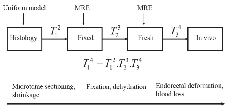
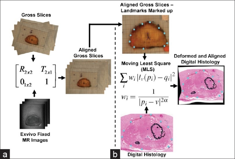
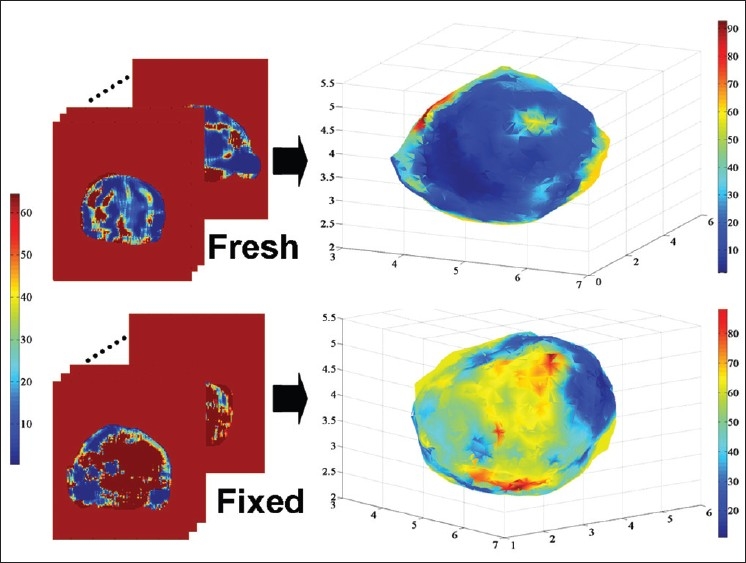
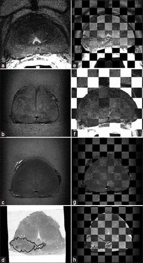
A biomechanical model-based deformable image registration incorporating specimen-specific changes in material properties is optimized and evaluated for correlating histology of clinical prostatectomy specimens with in vivo MRI. In this methodology, a three-step registration based on biomechanics calculates the transformations between histology and fixed, fixed and fresh, and fresh and in vivo states. A heterogeneous linear elastic material model is constructed based on magnetic resonance elastography (MRE) results. The ex vivo tissue MRE data provide specimen-specific information for the fresh and fixed tissue to account for the changes due to fixation. The accuracy of the algorithm was quantified by calculating the target registration error (TRE) by identifying naturally occurring anatomical points within the prostate in each image. TRE were improved with the deformable registration algorithm compared to rigid registration alone. The qualitative assessment also showed a good alignment between histology and MRI after the proposed deformable registration.
Keywords: Biomechanical models; correlative pathology; deformable registration; finite element model; magnetic resonance elastography.
Figures
Similar articles
-
SU-E-J-95: Towards Optimum Boundary Conditions for Biomechanical Model Based Deformable Registration Using Intensity Based Image Matching for Prostate Correlative Pathology.Med Phys. 2012 Jun;39(6Part7):3674. doi: 10.1118/1.4734931. Med Phys. 2012. PMID: 28519778
-
Validation of biomechanical deformable image registration in the abdomen, thorax, and pelvis in a commercial radiotherapy treatment planning system.Med Phys. 2017 Jul;44(7):3407-3417. doi: 10.1002/mp.12307. Epub 2017 Jun 1. Med Phys. 2017. PMID: 28453911 Free PMC article.
-
Development of a registration framework to validate MRI with histology for prostate focal therapy.Med Phys. 2015 Dec;42(12):7078-89. doi: 10.1118/1.4935343. Med Phys. 2015. PMID: 26632061
-
Accuracy of deformable image registration techniques for alignment of longitudinal cholangiocarcinoma CT images.Med Phys. 2020 Apr;47(4):1670-1679. doi: 10.1002/mp.14029. Epub 2020 Feb 12. Med Phys. 2020. PMID: 31958147 Free PMC article.
-
Comparison of physics-based deformable registration methods for image-guided neurosurgery.Front Digit Health. 2023 Dec 8;5:1283726. doi: 10.3389/fdgth.2023.1283726. eCollection 2023. Front Digit Health. 2023. PMID: 38144260 Free PMC article. Review.
Cited by
-
Effect of material property heterogeneity on biomechanical modeling of prostate under deformation.Phys Med Biol. 2015 Jan 7;60(1):195-209. doi: 10.1088/0031-9155/60/1/195. Epub 2014 Dec 9. Phys Med Biol. 2015. PMID: 25489840 Free PMC article.
-
Role of endorectal MR imaging and MR spectroscopic imaging in defining treatable intraprostatic tumor foci in prostate cancer: quantitative analysis of imaging contour compared to whole-mount histopathology.Radiother Oncol. 2014 Feb;110(2):303-8. doi: 10.1016/j.radonc.2013.12.003. Epub 2014 Jan 17. Radiother Oncol. 2014. PMID: 24444524 Free PMC article.
-
ProsRegNet: A deep learning framework for registration of MRI and histopathology images of the prostate.Med Image Anal. 2021 Feb;68:101919. doi: 10.1016/j.media.2020.101919. Epub 2020 Dec 17. Med Image Anal. 2021. PMID: 33385701 Free PMC article.
-
The mechanics of metastasis: insights from a computational model.PLoS One. 2012;7(9):e44281. doi: 10.1371/journal.pone.0044281. Epub 2012 Sep 28. PLoS One. 2012. PMID: 23028513 Free PMC article.
-
Enhancement pattern mapping technique for improving contrast-to-noise ratios and detectability of hepatobiliary tumors on multiphase computed tomography.Med Phys. 2020 Jan;47(1):64-74. doi: 10.1002/mp.13769. Epub 2019 Nov 19. Med Phys. 2020. PMID: 31449684 Free PMC article.
References
-
- Jaffray DA, Siewerdsen JH, Wong JW, Martinez AA. Flat-panel cone-beam computed to-mography for image-guided radiation therapy. Int J Radiat Oncol Biol Phys. 2002;53:1337–49. - PubMed
-
- Jeanneret-Sozzi W, Moeckli R, Valley JF, Zouhair A, Ozsahin EM, Mirimanoff RO. The reasons for discrepancies in target volume delineation. Strahlenther Onkol. 2006;182:450–7. - PubMed
-
- Teh BS, Bastasch MD, Wheeler TM, Mai W, Frolov A, Uhl BM, et al. IMRT for prostate cancer: defin-ing target volume based on correlated pathologic volume of disease. Int J Radiat Oncol Biol Phys. 2003;56:184–91. - PubMed
-
- Rueckert D, Sonoda LI, Hayes C, Hill DL, Leach MO, Hawkes DJ. Nonrigid registration using free-form deformations: Application to breast MR images. IEEE Trans Med Imaging. 2002;18:712–21. - PubMed
-
- Dauguet J, Delzescaux T, Condé F, Mangin JF, Ayache N, Hantraye P, et al. Three-dimensional reconstruction of stained histological slices and 3D non-linear registration with in vivo MRI for whole baboon brain. J Neurosci Methods. 2007;164:191–204. - PubMed
