

Basal ganglia circuits changes in Parkinson's disease patients
- PMID: 22813979
- PMCID: PMC4163196
- DOI: 10.1016/j.neulet.2012.07.012
Basal ganglia circuits changes in Parkinson's disease patients
Abstract
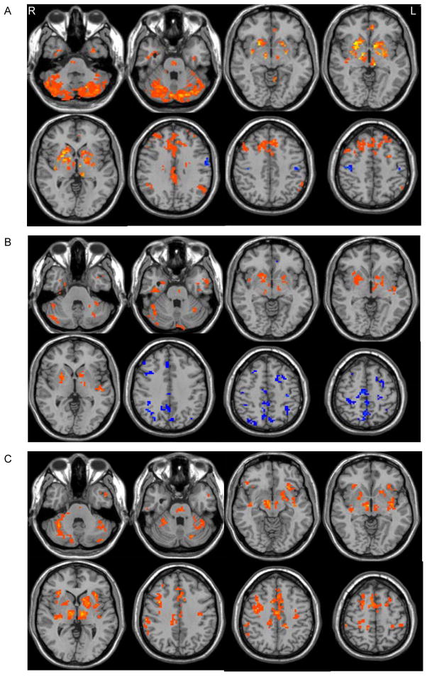
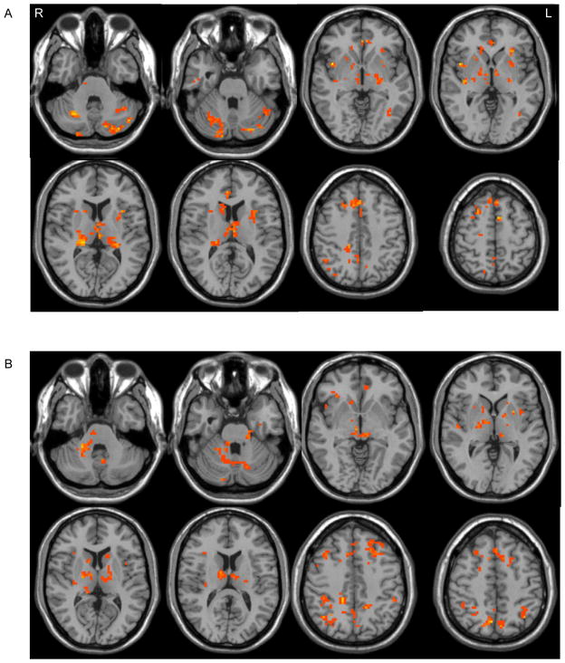
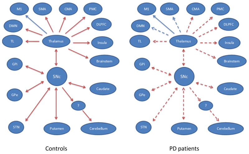
Functional changes in basal ganglia circuitry are responsible for the major clinical features of Parkinson's disease (PD). Current models of basal ganglia circuitry can only partially explain the cardinal symptoms in PD. We used functional MRI to investigate the causal connectivity of basal ganglia networks from the substantia nigra pars compacta (SNc) in PD in the movement and resting state. In controls, SNc activity predicted increased activity in the supplementary motor area, the default mode network, and dorsolateral prefrontal cortex, but, in patients, activity predicted decreases in the same structures. The SNc had decreased connectivity with the striatum, globus pallidus, subthalamic nucleus, thalamus, supplementary motor area, dorsolateral prefrontal cortex, insula, default mode network, temporal lobe, cerebellum, and pons in patients compared to controls. Levodopa administration partially normalized the pattern of connectivity. Our findings show how the dopaminergic system exerts influences on widespread brain networks, including motor and cognitive networks. The pattern of basal ganglia network connectivity is abnormal in PD secondary to dopamine depletion, and is more deviant in more severe disease. Use of functional MRI with network analysis appears to be a useful method to demonstrate basal ganglia pathways in vivo in human subjects.
Copyright © 2012 Elsevier Ireland Ltd. All rights reserved.
Figures
Similar articles
-
Beta-band oscillations in the supplementary motor cortex are modulated by levodopa and associated with functional activity in the basal ganglia.Neuroimage Clin. 2018 May 18;19:559-571. doi: 10.1016/j.nicl.2018.05.021. eCollection 2018. Neuroimage Clin. 2018. PMID: 29984164 Free PMC article.
-
Deep brain stimulation induced normalization of the human functional connectome in Parkinson's disease.Brain. 2019 Oct 1;142(10):3129-3143. doi: 10.1093/brain/awz239. Brain. 2019. PMID: 31412106
-
Cerebello-basal ganglia connectivity fingerprints related to motor/cognitive performance in Parkinson's disease.Parkinsonism Relat Disord. 2020 Nov;80:21-27. doi: 10.1016/j.parkreldis.2020.09.005. Epub 2020 Sep 7. Parkinsonism Relat Disord. 2020. PMID: 32932024
-
Functional changes of the basal ganglia circuitry in Parkinson's disease.Prog Neurobiol. 2000 Sep;62(1):63-88. doi: 10.1016/s0301-0082(99)00067-2. Prog Neurobiol. 2000. PMID: 10821982 Review.
-
Functional organization of the basal ganglia: therapeutic implications for Parkinson's disease.Mov Disord. 2008;23 Suppl 3:S548-59. doi: 10.1002/mds.22062. Mov Disord. 2008. PMID: 18781672 Review.
Cited by
-
Task-rest modulation of basal ganglia connectivity in mild to moderate Parkinson's disease.Brain Imaging Behav. 2015 Sep;9(3):619-38. doi: 10.1007/s11682-014-9317-9. Brain Imaging Behav. 2015. PMID: 25280970 Free PMC article.
-
Acute Modulation of Brain Connectivity in Parkinson Disease after Automatic Mechanical Peripheral Stimulation: A Pilot Study.PLoS One. 2015 Oct 15;10(10):e0137977. doi: 10.1371/journal.pone.0137977. eCollection 2015. PLoS One. 2015. PMID: 26469868 Free PMC article. Clinical Trial.
-
Altered subcortical emotional salience processing differentiates Parkinson's patients with and without psychotic symptoms.Neuroimage Clin. 2020;27:102277. doi: 10.1016/j.nicl.2020.102277. Epub 2020 May 30. Neuroimage Clin. 2020. PMID: 32540629 Free PMC article.
-
Cerebellar Contribution to Motor and Non-motor Functions in Parkinson's Disease: A Meta-Analysis of fMRI Findings.Front Neurol. 2020 Feb 27;11:127. doi: 10.3389/fneur.2020.00127. eCollection 2020. Front Neurol. 2020. PMID: 32174883 Free PMC article.
-
Aging with HIV-1 Infection: Motor Functions, Cognition, and Attention--A Comparison with Parkinson's Disease.Neuropsychol Rev. 2015 Dec;25(4):424-38. doi: 10.1007/s11065-015-9305-x. Epub 2015 Nov 17. Neuropsychol Rev. 2015. PMID: 26577508 Free PMC article. Review.
References
-
- Aarsland D, Andersen K, Larsen JP, Lolk A, Kragh-Sorensen P. Risk of dementia in Parkinson’s disease. A community-based, prospective study. Neurology. 2001;56:730–736. - PubMed
-
- Alexander GE, Crutcher MD. Functional architecture of basal ganglia circuits: neural substrates of parallel processing. Trends Neurosci. 1990;13:266–271. - PubMed
-
- Braak H, Del Tredici K, Rüba U, de Vos RAI, Jansen Steur ENH, Braak E. Staging of brain pathology related to sporadic Parkinson’s disease. Neurobiol Aging. 2003;24:197–211. - PubMed
-
- Chen G, Hamilton JP, Thomason ME, Gotlib IH, Saad ZS, Cox RW. Granger Causality via Vector Auto-Regression Tuned for FMRI Data Analysis. Proc Intl Soc Mag Reson Med. 2009;17:1718.
Publication types
MeSH terms
Grants and funding
LinkOut - more resources
Full Text Sources
Medical
Miscellaneous
