. 2012 Jul 31;109(31):12604-9.
doi: 10.1073/pnas.1204302109. Epub 2012 Jul 18.

Heterotrophic feeding as a newly identified survival strategy of the dinoflagellate Symbiodinium

Affiliations

Heterotrophic feeding as a newly identified survival strategy of the dinoflagellate Symbiodinium

Hae Jin Jeong et al. Proc Natl Acad Sci U S A. .

Abstract

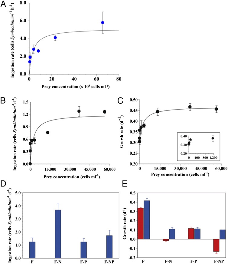
Survival of free-living and symbiotic dinoflagellates (Symbiodinium spp.) in coral reefs is critical to the maintenance of a healthy coral community. Most coral reefs exist in oligotrophic waters, and their survival strategy in such nutrient-depleted waters remains largely unknown. In this study, we found that two strains of Symbiodinium spp. cultured from the environment and acquired from the tissues of the coral Alveopora japonica had the ability to feed heterotrophically. Symbiodinium spp. fed on heterotrophic bacteria, cyanobacteria (Synechococcus spp.), and small microalgae in both nutrient-replete and nutrient-depleted conditions. Cultured free-living Symbiodinium spp. displayed no autotrophic growth under nitrogen-depleted conditions, but grew when provided with prey. Our results indicate that Symbiodinium spp.'s mixotrophic activity greatly increases their chance of survival and their population growth under nitrogen-depleted conditions, which tend to prevail in coral habitats. In particular, free-living Symbiodinium cells acquired considerable nitrogen from algal prey, comparable to or greater than the direct uptake of ammonium, nitrate, nitrite, or urea. In addition, free-living Symbiodinium spp. can be a sink for planktonic cyanobacteria (Synechococcus spp.) and remove substantial portions of Synechococcus populations from coral reef waters. Our discovery of Symbiodinium's feeding alters our conventional views of the survival strategies of photosynthetic Symbiodinium and corals.


Conflict of interest statement

The authors declare no conflict of interest.

Figures

Fig. 1.
Fig. 1.
Images of free-living Symbiodinium cells feeding on diverse prey. (AC) Micrographs of Symbiodinium sp. cells without added prey observed under a light microscope (A) and bacterium-sized fluorescent beads (1 μm in diameter) (B) and three beads (arrows) inside the protoplasm of Symbiodinium cells observed under an epifluorescent microscope (C). (DF) Micrographs of Symbiodinium sp. cells without added prey (D), fluorescently labeled bacteria (E), and three fluorescently labeled bacteria (arrows) inside the protoplasm of Symbiodinium cells observed under an epifluorescent microscope (F). (GI) Transmission electron microscope images showing Symbiodinium sp. with ingested Synechococcus sp. cells (arrows or inside a circle). Uningested Synechococcus cells were observed outside the predator cell (G and H). H is enlarged from G. (JO) Images of feeding by Symbiodinium cells on H. akashiwo prey. (J) Scanning electron micrograph of a Symbiodinium cell showing a peduncle (PE; feeding tube, arrow). (K) Light microscope image showing a Symbiodinium cell (Sm) deploying a tow filament and anchored on an H. akashiwo (Ha) (arrow). (L) A Symbiodinium cell (Sm) sucking materials from an H. akashiwo (Ha) cell through a peduncle (arrow). (M) A chloroplast of H. akashiwo (Ha) sucked by Symbiodinium cell (Sm) through a peduncle (arrow). (N) Several chloroplasts of the prey cell (greenish) were observed inside the protoplasm of the predator cell. (O) Two Symbiodinium cells (Sm 1 and Sm 2) simultaneously sucking materials (arrows) from an H. akashiwo (Ha) cell. (Scale bars: 5 μm for JO, 2 μm for DG, 1 μm for AC, and 0.5 μm for H and I.)
Fig. 2.
Fig. 2.
Transmission electron micrographs of free-living Symbiodinium feeding on H. akashiwo prey. (A) Micrograph showing a Symbiodinium cell (Sm) sucking materials from H. akashiwo (Ha) through a peduncle (PE). (B and DF) Images enlarged from A. The predator cell contains ingested prey chloroplasts (PCs) (B), which have less-dense thylakoids (T), most likely owing to semidigestion, compared with intact prey chloroplasts (C) and different from the chloroplasts of the predator (PDC) (D). (E) A prey chloroplast moving through a peduncle. (F) Enlarged image showing prey thylakoids (T) inside a prey chloroplast (PC). (Scale bars: 1 μm.)
Fig. 3.
Fig. 3.
Feeding processes of free-living Symbiodinium cells on H. akashiwo prey, observed under an inverted microscope and recorded by video microscopy. (A) An unfed H. akashiwo (Ha) cell containing many chloroplasts (20–35 chloroplasts per cell). A Symbiodinium cell (Sm) feeding on an H. akashiwo (Ha) prey after deploying a tow filament and then a peduncle (stage 1), as shown in Fig. 1 KM. (BD) A Symbiodinium cell (Sm) feeding on an H. akashiwo (Ha) prey with spinning (stage 2). In stage 2, several prey chloroplasts were sucked by the predator. (EG) A Symbiodinium cell (Sm) feeding on an H. akashiwo prey (Ha) after it had stopped rotating (stage 3). In stage 3, additional prey chloroplasts (blue and red arrows indicate different chloroplasts) were sucked by the predator again, after which the prey chloroplasts collapsed and burst, most likely owing to large empty spaces where prey chloroplasts occurred previously. Approximately 8–10 chloroplasts were observed within the burst prey cell. (H and I) Additional prey chloroplasts (yellow arrows) were sucked again by the predator from the burst prey cell (stage 4). At the end of stage 4, several empty (i.e., completely digested, white arrows) and one or two thylakoid-retained (green arrows) chloroplasts were observed inside the prey cell (I). (Scale bars: 5 μm.)
Fig. 4.
Fig. 4.
Feeding rates of free-living Symbiodinium sp. on Synechococcus sp. and H.akashiwo prey. (A) Ingestion rate of Symbiodinium on Synechococcus as a function of the initial prey concentration (x, cells mL−1). Symbols represent treatment mean ±1 SE. The curves are fitted to the Michaelis–Menten equations using all of the treatments. Ingestion rate: (cells Symbiodinium−1 h−1) = 5.3 [x/(3.9 × 105 + x)], R2 = 0.581. (B and C) Ingestion (B) and growth rates (C) of Symbiodinium sp. on H. akashiwo as a function of the mean prey concentration (x, cells mL−1). Ingestion rate: (cells Symbiodinium−1 d−1) = 1.2 [x/(2,280 + x)]; R2 = 0.838. Growth rate: (d−1) = 0.467 [(x + 1,720)/(880 + (x + 1,720))]; R2 = 0.608. (D and E) Effects of inorganic nutrients on feeding rates of free-living Symbiodinium sp. Ingestion (D) and growth rates (E) of Symbiodinium sp. on H. akashiwo under different nutrient conditions; Red bars indicate no added prey; blue bars, mean prey concentrations = 15,200–19,600 cells mL−1. F, f/2 medium (see Materials and Methods for concentrations); F-N, N-depleted f/2 medium; F-P, P-depleted f/2 medium; F-NP, N- and P- depleted f/2 medium. Symbols represent treatment mean ±1 SE.

References

    1. Hughes TP, et al. Climate change, human impacts, and the resilience of coral reefs. Science. 2003;301:929–933. - PubMed
    1. Tang YZ, Koch F, Gobler CJ. Most harmful algal bloom species are vitamin B1 and B12 auxotrophs. Proc Natl Acad Sci USA. 2010;107:20756–20761. - PMC - PubMed
    1. Mumby PJ, Hastings A, Edwards HJ. Thresholds and the resilience of Caribbean coral reefs. Nature. 2007;450:98–101. - PubMed
    1. Pennisi E. Reefs in trouble: Spawning for a better life. Science. 2007;318:1712–1717. - PubMed
    1. Maina J, McClanahan TR, Venus V, Ateweberhan M, Madin J. Global gradients of coral exposure to environmental stresses and implications for local management. PLoS ONE. 2011;6:e23064. - PMC - PubMed

Publication types

Associated data

LinkOut - more resources