Comparative Study
. 2012 Oct;117(4):836-46.
doi: 10.1097/ALN.0b013e3182655e73.

Intralipid, a clinically safe compound, protects the heart against ischemia-reperfusion injury more efficiently than cyclosporine-A

Affiliations
Comparative Study

Intralipid, a clinically safe compound, protects the heart against ischemia-reperfusion injury more efficiently than cyclosporine-A

Jingyuan Li et al. Anesthesiology. 2012 Oct.

Abstract

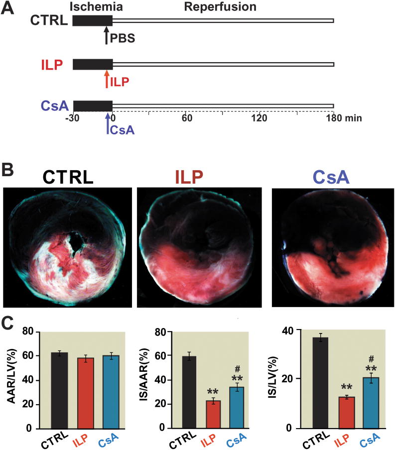
Background: We have recently shown that postischemic administration of intralipid protects the heart against ischemia-reperfusion injury. Here we compared the cardioprotective effects of intralipid with cyclosporine-A, a potent inhibitor of the mitochondrial permeability transition pore opening.

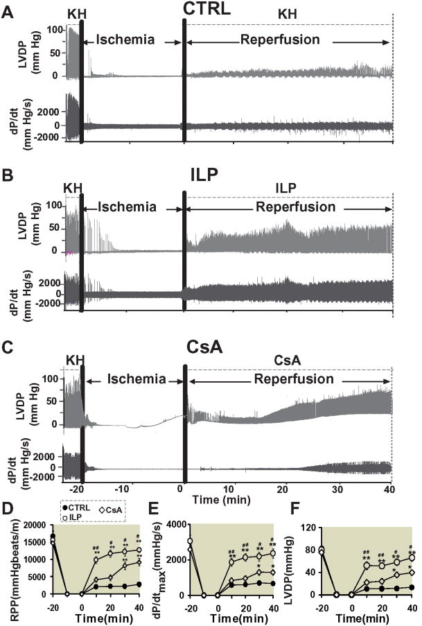
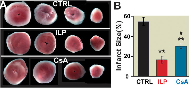
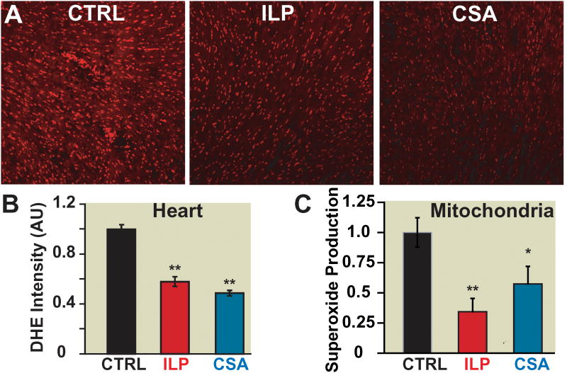
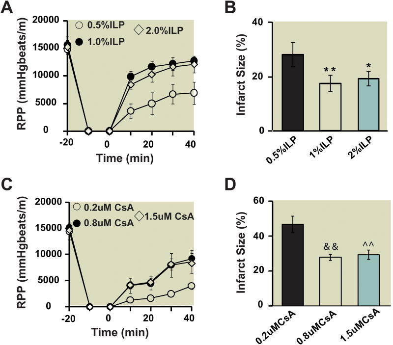
Methods: In vivo rat hearts or isolated Langendorff-perfused mouse hearts were subjected to ischemia followed by reperfusion with intralipid (0.5%, 1% and 2% ex-vivo, and 20% in vivo), cyclosporine-A (0.2 μM, 0.8 μM, and 1.5 μM ex- vivo and 10 mg/kg in vivo), or vehicle. The hemodynamic function, infarct size, calcium retention capacity, mitochondrial superoxide production, and phosphorylation levels of protein kinase B (Akt)/glycogen synthase kinase-3β (GSK-3β) were measured. The values are mean ± SEM.

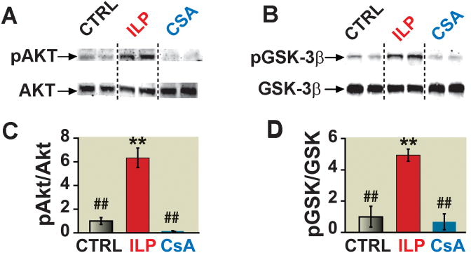
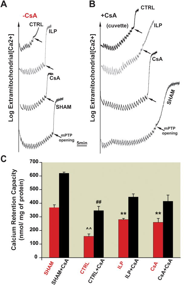
Results: Administration of intralipid at reperfusion significantly reduced myocardial infarct size compared with cyclosporine-A in vivo (infarct size/area at risk)%: 22.9 ± 2.5% vs. 35.2 ± 3.5%; P = 0.030, n = 7/group). Postischemic administration of intralipid at its optimal dose (1%) was more effective than cyclosporine-A (0.8 μM) in protecting the ex vivo heart against ischemia-reperfusion injury, as the rate pressure product at the end of reperfusion was significantly higher (mmHg · beats/min: 12,740 ± 675 [n = 7] vs. 9,203 ± 10,781 [n = 5], P = 0.024), and the infarct size was markedly smaller (17.3 ± 2.9 [n = 7] vs. 29.2 ± 2.7 [n = 5], P = 0.014). Intralipid was as efficient as cyclosporine-A in inhibiting the mitochondrial permeability transition pore opening (calcium retention capacity = 280 ± 8.2 vs. 260.3 ± 2.9 nmol/mg mitochondria protein in cyclosporine-A, P = 0.454, n = 6) and in reducing cardiac mitochondrial superoxide production. Unlike intralipid, which increased phosphorylation of Akt (6-fold) and GSK-3β (5-fold), cyclosporine-A had no effect on the activation of these prosurvival kinases.

Conclusions: Although intralipid inhibits the opening of the mitochondrial permeability transition pore as efficiently as cyclosporine-A, intralipid is more effective in reducing the infarct size and improving the cardiac functional recovery.


Figures

Figure 1
Figure 1. Intralipid reduces the infarct size more efficiently than Cyclosporine-A in the in vivo ischemia/reperfusionrat model
A. The left coronary artery was occluded for 30 minutes followed by 3 hr of reperfusion. One single IV bolus of 20% Intralipid (5ml/kg body weight, intralipid group), Cyclosporine-A(10mg/kg body weight, cyclosporine-A group) and phosphate buffered saline (control group, CTRL) was administered 5 min before reperfusion. B. representativetriphenyltetrazolium chloride-stained heart slices from from control, intralipid(ILP) and cyclosporine-A(CsA) group. The white area represents show infracted area (Corresponding drawings), blue area shows noneinfarct area red and white areas show risk area. C. Percentage of area at risk (AAR) divided by left ventricle (LV) (B), infarct size (IS) divided by AAR (C), and infarct size (IS) divided by left ventricle in CTRL(n=7), intralipid group(n=7) and cyclosporine-A group (n=7). **p<0.001 vs. CTRL, #p=0.030 vs. CsA.
Figure 2
Figure 2. Dose response of intralipid and cyclosporine-A in ex vivo ischemia/reperfusionmouse model
A. Rate pressure product (RPP) as a function of time in 0.5%, 1% and 2% intralipid (n=7). B. The area of necrosis as the percentage of total ventricular area in 0.5%, 1% and 2% intralipidgroup(ILP). C. Rate pressure product as a function of time in 0.2μM, 0.8μM and 1.5μM cyclosporine-A(CsA)( n=5). D. The area of necrosis as the percentage of total ventricular area in control group(CTRL), 0.5%, 1% and 2% intralipid group. **p=0.004, 1% ILP vs. 0.5% ILP; *p=0.022, 2%ILP vs. 0.5% ILP; &&p=0.008, 0.8μMCsA vs. 0.2μM CsA; ^^p=0.009, 1.5μM CsA vs. 0.2μM CsA.
Figure 3
Figure 3. Administration of intralipid at reperfusion improves heart functional recovery against reperfusion injury more efficiently than cyclosporine-A
Representatives of the left ventricular developed pressure (LVDP) and dP/dtMax and dP/dtMin as a function of time in control group(CTRL)(A), 1%intralipid(ILP) (B) and 0.8μM cyclosporine-A(CsA)(C). Rate pressure product (D), dP/dtMax(E) and LVDP (F) as a function of time in CTRL (filled circles, n=7), ILP (open circles, n=7) and cyclosporine-A group (CsA, dimanods, n=5). *p<0.05 and **p< 0.001 vs. CTRL, #p< 0.05, ##p< 0.01 vs.CsA.
Figure 4
Figure 4. The infarct size is significantly smaller in intralipid than cyclosporine-A group
Four slices of the same heart after 2,3,5-triphenyltetrazolium chloride (TTC) staining in control group(CTRL)(n=7) (A), 1% intralipid group(ILP) (n=7) (B) and 0.8 μM cyclosporine-A group(n=5) (C). The white area represents the infarct zone and the red shows the viable area. E. The area of necrosis as the percentage of total ventricular area in CTRL (black), intralipid (red) and cyclosporine-A group(blue). **p< 0.001 vs. CTRL, #p=0.014 vs. ILP.
Figure 5
Figure 5. Postischemic treatment of intralipid increases mitochondrial CRC after I/R as cyclosporine-A in a CypD-dependent manner. A, B
Typical recordings of the mitochondrial permeability transition pore opening in isolated mitochondria from control group (CTRL), 1% intralipid (ILP) and 1.5 μM cyclosporine-A groups (CsA) subjected to 20 min of global ischemia followed by 10 min of reperfusion as well as sham hearts before (A) and after addition of 1.5μM cyclosporine-A directly in the cuvette (B). C. CRC in the absence of cyclosporine-A (red bars) and after addition of cyclosporine-A in the cuvette (black bars). **p<0.001 vs. CTRL; ##p<0.001 vs. sham+ cyclosporine-A group; ^^p<0.001 vs. sham (n=6).
Figure 6
Figure 6. Post-ischemic treatment of intralipid and cyclosporine-A decreases cardiac reactive oxygen species generation as well as mitochondrial superoxide production
A. Representative dihidroethidium staining of transverse heart sections subjected to 20min of global ischemia followed by 5min reperfusion with phosphate buffered saline (control group, CTRL) (black), 1% intralipidgroup(ILP) (red) and 1.5 μM cyclosporine-A group(CsA)(blue). B. Fluorescence quantification of dihydroethidium (DHE) staining: average intensity represents area×fluorescence intensity and is normalized to CTRL (**p<0.001 vs. CTRL, n=3). C. Superoxide production in isolated mitochondria using electron spin resonance in CTRL (black), 1% intralipid (red) and 1.5 μMcyclosporine-A group(blue). **p=0.005 ILP vs. CTRL; *p=0.041 CsA vs. CTRL (n=4).
Figure 7
Figure 7. The lack of involvement of Akt/GSK pathways in cyclosporine-A-induced protection
A, B. representative immunoblots of pAkt and total Akt (A), and pGSK-3β and total GSK-3β (C) in heart homogenates subjected to I/R from control group(CTRL), 1% intralipid, or 1.5 μM cyclosporine-A group. C, D. Western blot analysis of pAkt protein to total Akt (B) and pGSK-3β to total GSK-3b (D) ratios in CTRL (black bars), intralipid group (ILP, white bars) and cyclosporine-A group(gray bars). **p<0.001 vs. CTRL; ##p<0.001 vs. ILP (n=4−6/group).

References

    1. Murphy E, Steenbergen C. Mechanisms underlying acute protection from cardiac ischemia-reperfusion injury. Physiol Rev. 2008;88:581–609. - PMC - PubMed
    1. Gorczynski RJ. Basic pharmacology of esmolol. Am J Cardiol. 1985;56:3F–13F. - PubMed
    1. Sugimoto S, Puddu PE, Monti F, Schiariti M, Campa PP, Marino B. Pretreatment with the adenosine triphosphate-sensitive potassium channel opener nicorandil and improved myocardial protection during high-potassium cardioplegic hypoxia. J Thorac Cardiovasc Surg. 1994;108:455–66. - PubMed
    1. Dorman BH, Hebbar L, Zellner JL, New RB, Houck WV, Acsell J, Nettles C, Hendrick JW, Sampson AP, Mukherjee R, Spinale FG. ATP-sensitive potassium channel activation before cardioplegia. Effects on ventricular and myocyte function. Circulation. 1998;98(II):176–83. - PubMed
    1. Cour M, Gomez L, Mewton N, Ovize M, Argaud L. Postconditioning: From the bench to bedside. J Cardiovasc Pharmacol Ther. 2011;16:117–30. - PubMed

Publication types

MeSH terms