. 2012 Oct;50(10):775-82.
doi: 10.1002/dvg.22049. Epub 2012 Aug 16.

Generation and live imaging of an endogenous Cdx2 reporter mouse line

Affiliations

Generation and live imaging of an endogenous Cdx2 reporter mouse line

Katie McDole et al. Genesis. 2012 Oct.

Abstract

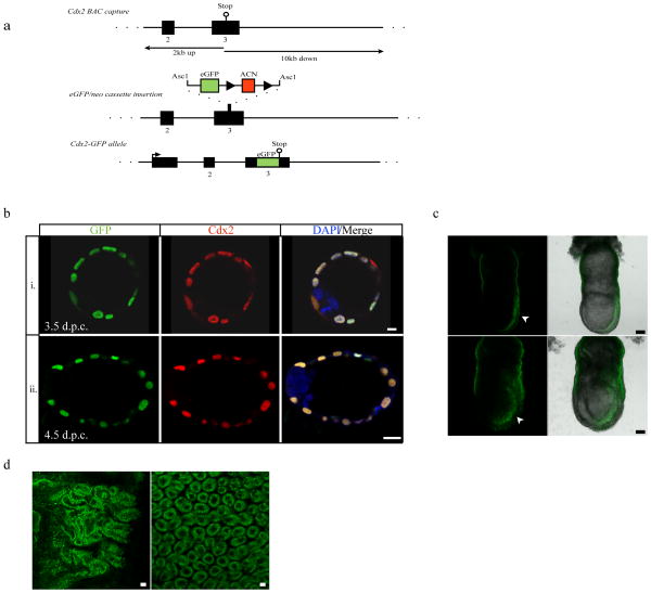
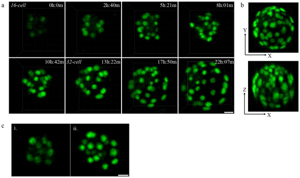
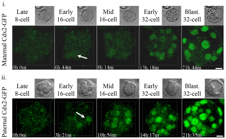
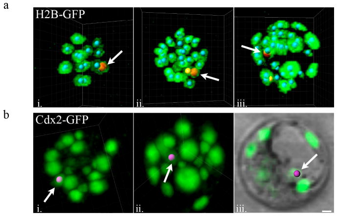
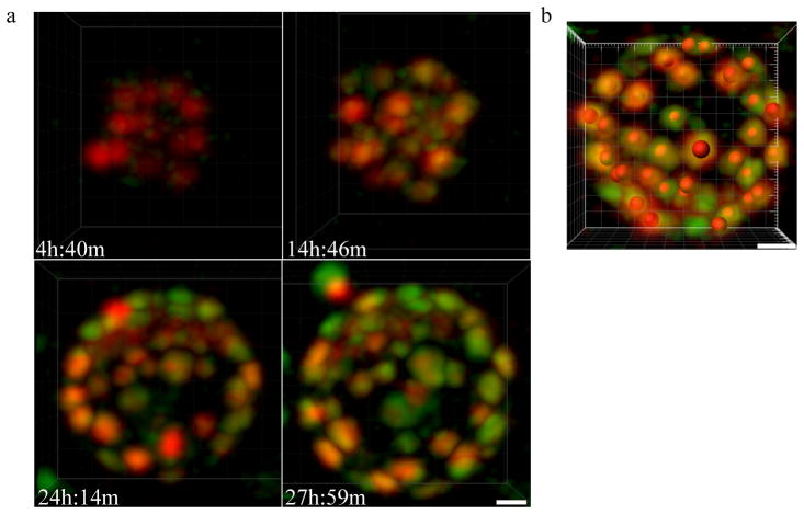
To understand cell fate specification and maintenance during development, it is essential to visualize both lineage markers and cell behaviors in real time using endogenous markers to report cell fate. We have generated a reporter line in which eGFP is fused to the endogenous locus of Cdx2, a transcription factor essential for trophectoderm specification, allowing us to visualize cell fate decisions in the preimplantation mouse embryo. We used two-photon laser scanning microscopy to visualize expression of the endogenous Cdx2 fusion protein and show that Cdx2 undergoes phases of upregulation. Additionally, we show that as late as the 32-cell stage, outer trophectoderm cells may change their fates by migrating inward and losing Cdx2 expression. Furthermore, the tools and techniques we report allow for dual-colored imaging, which will greatly facilitate the study of not only preimplantation development, but later stages of development and tissues where Cdx2 plays an important role.


Figures

Figure 1
Figure 1
Cdx2-GFP fusion allele expressed from the endogenous promoter. (a) Recombineering strategy to create a targeted Cdx2-GFP fusion protein by knocking-in GFP using targeted Asc1 restriction sites to introduce the reporter/selection cassette. Mice either heterozygous or homozygous for the Cdx2-GFP fusion allele are viable and fertile. (b) GFP expression in the Cdx2-GFP heterozygous embryos co-localizes with the endogenous Cdx2 as determined by immunofluorescence. GFP survives fixation and is visible at pre- and peri-implantation stages. i) 3.5 dpc. ii) 4.5 dpc. Scale bars, 10 μm (top), 20 μm (bottom). (c) Cdx2-GFP expression in two (top and bottom) live 7.0–7.5 d.p.c. heterozygous embryos. GFP expression can be found in the posterior end in the primitive streak (arrow heads). Scale bars, 70 μm (top), 100 μm (bottom). (d) Cdx2-GFP expression in live small intestine from heterozygous 5-week old female mice. GFP expression is found throughout the epithelial layer (left) and villi (right). Scale bars, 20 μm.
Figure 2
Figure 2
Imaging of endogenous Cdx2 expression live. (a) Cdx2-GFP expression from the 16-cell stage to the 64-cell stage. Still frames taken from a time-lapse movie of a single embryo using TPLSM show Cdx2-GFP expression beginning at the early to mid 16-cell stage, and is gradually up-regulated over time. Embryos are viable and develop with minimal or no delay under these conditions. Scale bar, 20 μm. (b) Two-photon microscopy provides full-thickness resolution of an expanded 64-cell stage blastocyst in both XY (top) and XZ (bottom) planes. (c) Increase of Cdx2-GFP expression following 16-cell to 32-cell division. Cdx2-GFP is noticeably brighter immediately after dividing from the 16-cell stage (i.) to the 32-cell stage (ii.). Scale bar, 20 μm.
Figure 3
Figure 3
Expression of Cdx2-GFP from maternal (Cdx2-GFP mother and CD1 WT father) (i.) or paternal (CD1 WT mother and Cdx2-GFP father) (ii.) alleles begins at the 16-cell stage (arrows indicate start of expression). Scale bar, 15 μm. Movies were obtained on two different days using different imaging settings. Maternal Cdx2-GFP was imaged using a conventional scanner, Paternal Cdx2-GFP was imaged under similar conditions using a resonant scanner.
Figure 4
Figure 4
Transient-outer cells expressing Cdx2-GFP migrate inward during cavitation. (a) Still frames taken from time-lapse movies using transgenic H2B-GFP expressing embryos. Nucleus marked with red “spot” and denoted by arrow marks a transient outer-cell that moves inward during cavitation. Nucleus marked with yellow “spot” is the outer-cell sibling of the red-labeled transient outer-cell after the 16-cell to 32-cell division (i. 16-cell stage, ii. 32-cell stage, iii. 32-cell blastocyst stage) (b) Cdx2 positive outer-cell at the 32 cell stage (i., nucleus labeled with pink “spot”) moves inward during cavitation (ii.) and shortly thereafter in the cavitated blastocyst resides in the ICM facing the blastocoel cavity (iii.). Scale bar, 10 μm.
Figure 5
Figure 5
Dual-colored imaging of a mouse embryo using TPLSM. (a) Still images from a time-lapse movie of an embryo expressing both H2B-mCherry and Cdx2-GFP. Embryos were imaged using 740 nm (mCherry) and 890 nm (GFP) excitation wavelengths and a resonant scanner. Scale bar, 15 μm. Gaussian filtering was performed in IMARIS to eliminate noise created by use of the resonant scanner. (b) A single frame from a movie of H2B-mCherry and Cdx2-GFP expressing embryos at the 64-cell stage is shown. Red spots mark nuclear positions representing H2B-mCherry positive nuclei. Scale bar, 20 μm.

References

    1. Deschamps J, van Nes J. Developmental regulation of the Hox genes during axial morphogenesis in the mouse. Development. 2005;132:2931–2942. - PubMed
    1. Grainger S, Savory JG, Lohnes D. Cdx2 regulates patterning of the intestinal epithelium. Developmental biology. 2010;339:155–165. - PubMed
    1. McDole K, Xiong Y, Iglesias PA, Zheng Y. Lineage mapping the pre-implantation mouse embryo by two-photon microscopy, new insights into the segregation of cell fates. Developmental biology. 2011;355:239–249. - PMC - PubMed
    1. Morris SA, Teo RT, Li H, Robson P, Glover DM, Zernicka-Goetz M. Origin and formation of the first two distinct cell types of the inner cell mass in the mouse embryo. Proc Natl Acad Sci U S A. 2010;107:6364–6369. - PMC - PubMed
    1. Moskaluk CA, Zhang H, Powell SM, Cerilli LA, Hampton GM, Frierson HF., Jr Cdx2 protein expression in normal and malignant human tissues: an immunohistochemical survey using tissue microarrays. Modern pathology: an official journal of the United States and Canadian Academy of Pathology, Inc. 2003;16:913–919. - PubMed

Publication types

LinkOut - more resources