

Review
doi: 10.1101/cshperspect.a011544.
The mechanism of eukaryotic translation initiation: new insights and challenges
Affiliations
- PMID: 22815232
- PMCID: PMC3475172
- DOI: 10.1101/cshperspect.a011544
Item in Clipboard
Review
The mechanism of eukaryotic translation initiation: new insights and challenges
Cold Spring Harb Perspect Biol.
.
Abstract
Translation initiation in eukaryotes is a highly regulated and complex stage of gene expression. It requires the action of at least 12 initiation factors, many of which are known to be the targets of regulatory pathways. Here we review our current understanding of the molecular mechanics of eukaryotic translation initiation, focusing on recent breakthroughs from in vitro and in vivo studies. We also identify important unanswered questions that will require new ideas and techniques to solve.
Figures
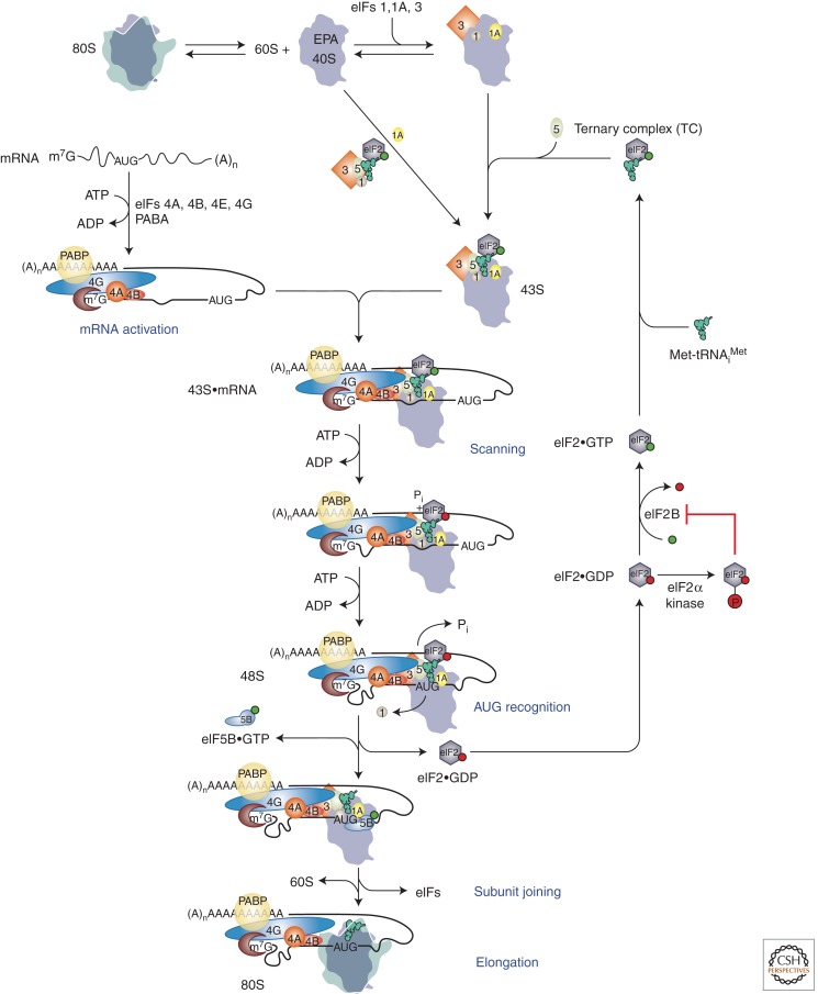
Model of canonical eukaryotic translation initiation pathway. The pathway is shown as a series of discrete steps starting with dissociation of 80S ribosomes into subunits. Binding of factors is depicted both as a single step via the multifactor complex and as two separate steps, with eIFs 1, 1A, and 3 binding first followed by binding of ternary complex and eIF5. The resulting 43S preinitiation complex (PIC) is then loaded onto an activated mRNP near the 5′ cap. Subsequent scanning of the mRNA allows recognition of the start codon, which triggers downstream steps in the pathway including eIF1 release from the PIC, Pi release from eIF2, and conversion to the closed, scanning-arrested state of the complex. eIF2·GDP released after subunit joining is recycled back to eIF2·GTP by the exchange factor eIF2B. eIF5B in its GTP-bound form promotes joining of the 60S subunit to the preinitiation complex, which triggers release of eIF5B·GDP and eIF1A to form the final 80S initiation complex, which can begin the elongation phase of protein synthesis. Throughout, GTP is depicted as a green ball and GDP as a red ball. (Modified from Hinnebusch 2011; reproduced, with permission, from the author.)
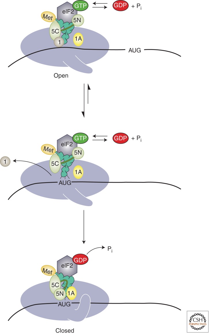
Model of structural rearrangements in the PIC accompanying start codon recognition. (Top) Before start codon recognition, the PIC exists in an open conformation, promoted by eIF1 and eIF1A, which is capable of scanning the mRNA. (Middle) Base pairing between the anticodon of the initiator tRNA and the start codon promotes movement of the tRNA from the Pout to Pin states and release of eIF1 from the complex. (Bottom) Ejection of eIF1, in turn, triggers release of Pi from eIF2, converting it to its GDP-bound form. Because eIF1 stabilizes the open state of the PIC, its departure also results in conversion of the complex to the closed, scanning-arrested conformation (shown as the closure of a latch on the mRNA entry site). Release of eIF1 is promoted by eIF5, possibly by competition between one of eIF5’s domains (depicted here as the amino-terminal domain; 5N) and eIF1 for the same binding site in the PIC. Start codon recognition also induces an interaction between eIF1A and eIF5, which further stabilizes the closed state of the complex. (Modified from Hinnebusch 2011; reproduced, with permission.)
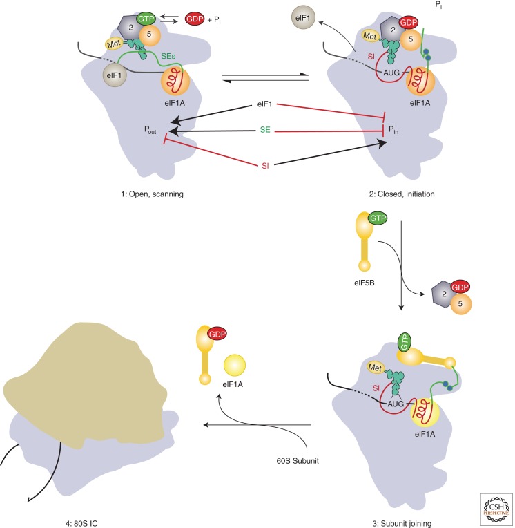
Model of the roles of eIF1A’s amino- and carboxy-terminal tails in mediating start codon recognition and later steps in eukaryotic translation initiation. Before start codon recognition (complex 1) the amino- and carboxy-terminal tails (CTTs; shown in red and green, respectively) are both in the P site of the 40S subunit. On start codon recognition (complex 2), the initiator tRNA moves from the Pout to Pin state, causing both eIF1 and the CTT of eIF1A to be ejected from the P site. eIF1 stabilizes the open conformation of the PIC and the Pout state of the initiator tRNA. The scanning enhancer (SE) elements in the CTT of eIF1A (shown as blue balls) stabilize the open state of the PIC relative to the closed state. Conversely, the scanning inhibitor (SI) element in eIF1A’s NTT destabilizes the open state, thus promoting closed complex formation. The CTT of eIF1A may interact directly with eIF5 after start codon recognition, and it is hypothesized that this interaction triggers Pi release from eIF2. After Pi release and dissociation of eIF2·GDP and eIF5 from the PIC (complex 3), the CTT of eIF1A is free to interact with eIF5B·GTP, recruiting it to the complex and promoting subunit joining. Release of eIF5B·GDP and eIF1A from the resulting 80S IC produces the final translation-competent ribosome, poised at the start codon to commence decoding of the mRNA. (Modified from Hinnebusch 2011; reproduced, with permission.)
References
-
- Acker MG, Shin BS, Dever TE, Lorsch JR 2006. Interaction between eukaryotic initiation factors 1A and 5B is required for efficient ribosomal subunit joining. J Biol Chem 281: 8469–8475 - PubMed
Publication types
MeSH terms
Substances
LinkOut - more resources
Full Text Sources
Other Literature Sources