. 2012 Sep 14;287(38):31866-76.
doi: 10.1074/jbc.M112.372169. Epub 2012 Jul 19.

The binding site of the V-ATPase inhibitor apicularen is in the vicinity of those for bafilomycin and archazolid

Affiliations

The binding site of the V-ATPase inhibitor apicularen is in the vicinity of those for bafilomycin and archazolid

Christin Osteresch et al. J Biol Chem. .

Abstract

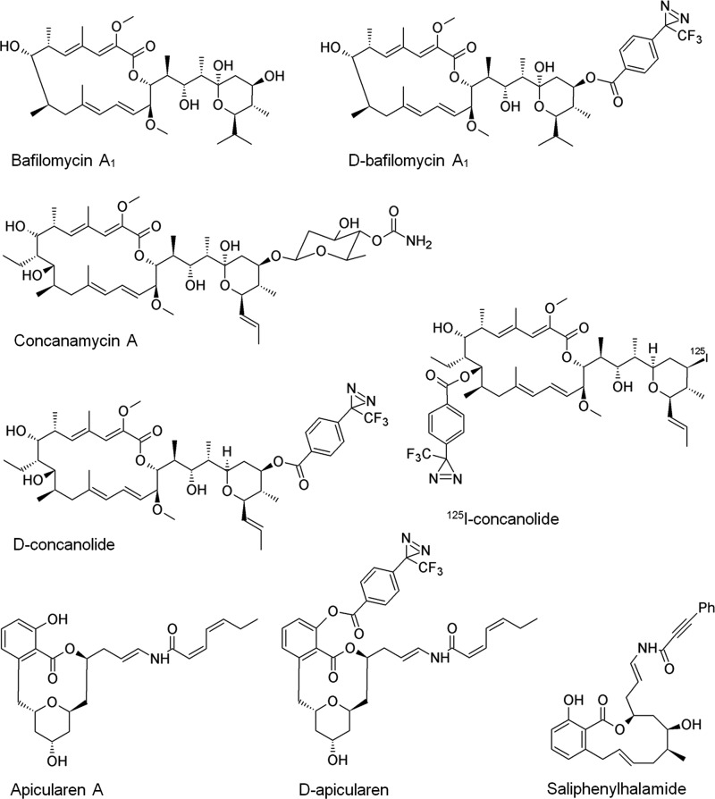
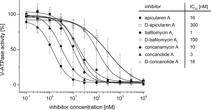
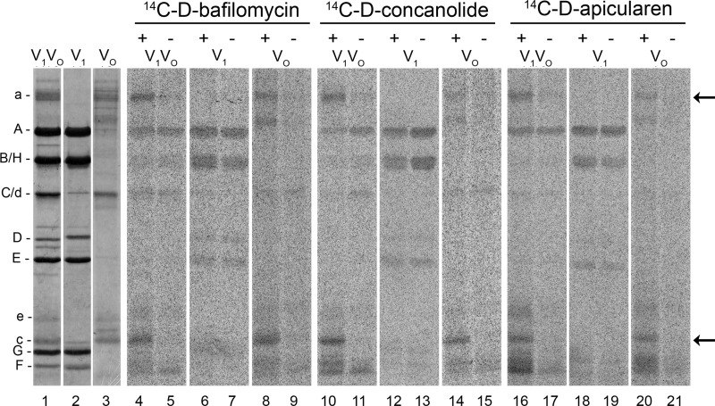
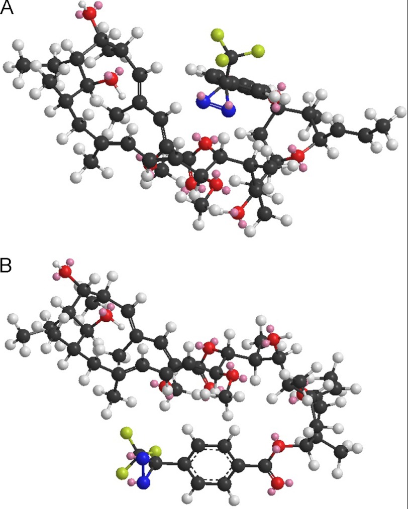
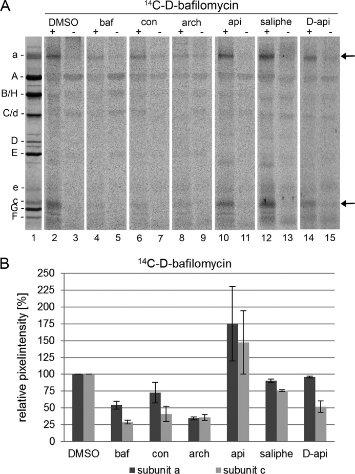
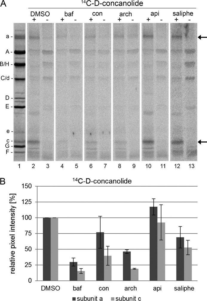
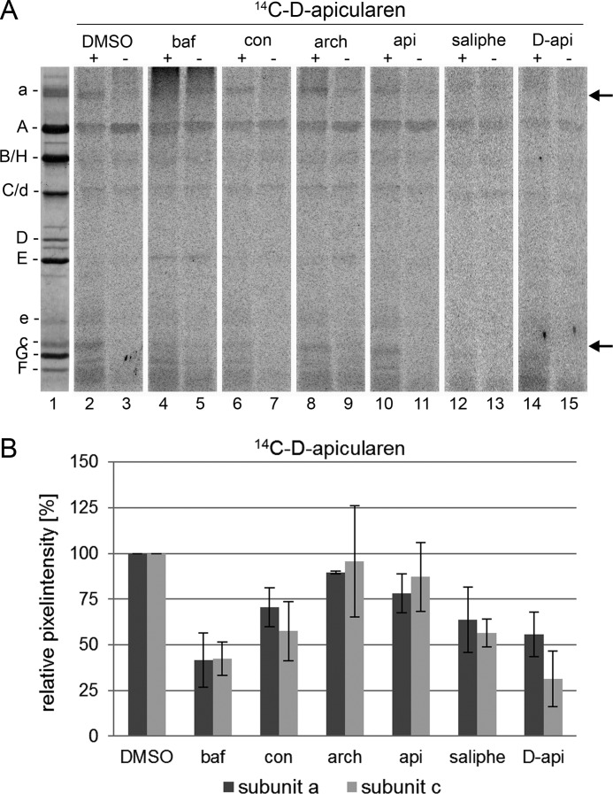
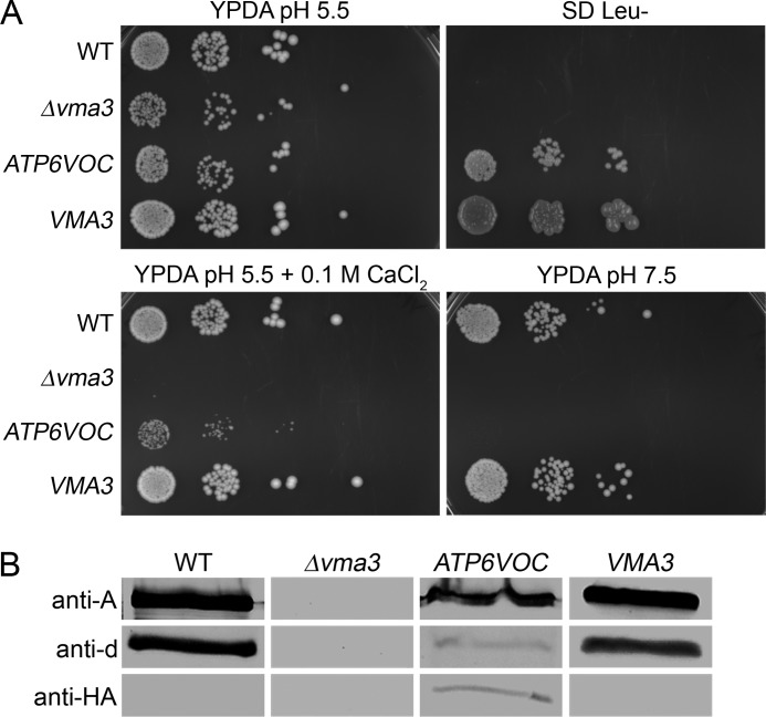
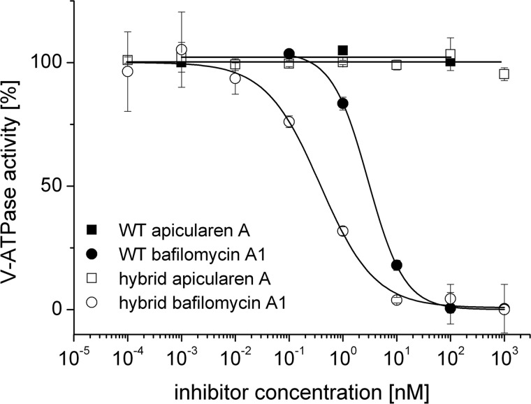
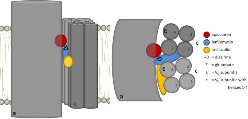
The investigation of V-ATPases as potential therapeutic drug targets and hence of their specific inhibitors is a promising approach in osteoporosis and cancer treatment because the occurrence of these diseases is interrelated to the function of the V-ATPase. Apicularen belongs to the novel inhibitor family of the benzolactone enamides, which are highly potent but feature the unique characteristic of not inhibiting V-ATPases from fungal sources. In this study we specify, for the first time, the binding site of apicularen within the membrane spanning V(O) complex. By photoaffinity labeling using derivatives of apicularen and of the plecomacrolides bafilomycin and concanamycin, each coupled to (14)C-labeled 4-(3-trifluoromethyldiazirin-3-yl)benzoic acid, we verified that apicularen binds at the interface of the V(O) subunits a and c. The binding site is in the vicinity to those of the plecomacrolides and of the archazolids, a third family of V-ATPase inhibitors. Expression of subunit c homologues from Homo sapiens and Manduca sexta, both species sensitive to benzolactone enamides, in a Saccharomyces cerevisiae strain lacking the corresponding intrinsic gene did not transfer this sensitivity to yeast. Therefore, the binding site of benzolactone enamides cannot be formed exclusively by subunit c. Apparently, subunit a substantially contributes to the binding of the benzolactone enamides.


Figures

FIGURE 1.
FIGURE 1.
Structures of the PAL inhibitor derivatives D-bafilomycin, D-concanolide, 125I-concanolide, D-apicularen, saliphenylhalamide, and parent compounds.
FIGURE 2.
FIGURE 2.
Inhibition of the ATPase activity of the M. sexta V1VO holoenzyme by bafilomycin, concanamycin, apicularen, and their derivatives. Values represent the means ± S.D. of experiments with three different preparations. The specific enzyme activity of the controls without inhibitors was 1.6 ± 0.4 μmol·mg−1·min−1.
FIGURE 3.
FIGURE 3.
Photoaffinity labeling of M. sexta V1VO holoenzyme, V1 complex, and VO complex with the 14C-labeled derivatives of D-bafilomycin, D-concanolide, and D-apicularen. For the labeling assays, 30 μg of V1VO holoenzyme, 20 μg of V1 complex, or 10 μg of VO complex were first incubated for 5 min at 25 °C with 52 μm 14C-D-bafilomycin, 52 μm 14C-D-concanolide, or 100 μm 14C-D-apicularen, respectively. Before the samples were exposed to UV light (366 nm) for 1 min (+) or kept in the dark (−), 1 mm Mg-ATP was added. Afterward, the protein subunits were separated by SDS-PAGE, stained with Coomassie Blue, and exposed to a phosphor screen. Lanes 1–3, typical staining with Coomassie Blue; lanes 4–21, readout of the phosphor screen.
FIGURE 4.
FIGURE 4.
Minimum energy calculation for D-concanolide. For 21-deoxyconcanolide A, the space of the connected diazirinyl moiety is placed preferably at the east hemisphere. The length of the benzoyl-diazirinyl label can be calculated to 6.4 Å and its distance from the macrolactone ring to 5.4 Å (A). Minimum energy calculations suggest that the diazirinyl group is situated preferably above and below the macrolactone ring very close to the molecule (A and B). A, benzoyl diazirinyl system is on the other side of the carbonyl group of the macrolactone ester. B, benzoyl diazirinyl system is on the same side of the carbonyl group of the macrolactone ester.
FIGURE 5.
FIGURE 5.
Western blot analysis of the V-ATPase with 14C-D-apicularen. For the identification of the 14C-D-apicularen-labeled band with the approximate molecular mass of subunit e 30 μg of the V1VO holoenzyme were incubated with 100 μm 14C-D-apicularen, and before the samples were exposed to UV light (366 nm) or kept in the dark, 1 mm Mg-ATP was added. Afterward, the protein subunits were separated by SDS-PAGE. Lane 1, staining of the SDS-gel with Coomassie Blue; lane 2, staining of the proteins with Ponceau S after electro-transfer onto a nitrocellulose membrane; lane 3, immunostaining with monoclonal anti-A (221-9) and anti-e (224-3) antibodies; lane 4, autoradiography of the irradiated sample; lane 5, autoradiography of the nonirradiated sample.
FIGURE 6.
FIGURE 6.
Photoaffinity labeling of the V-ATPase with 14C-D-bafilomycin after preincubation with different inhibitors. Samples with 30 μg of V1VO holoenzyme were preincubated for 5 min at 25 °C with DMSO, 625 μm bafilomycin (baf), concanamycin (con), archazolid (arch), apicularen (api), or saliphenylhalamide (saliphe), respectively. Then the samples were incubated for 5 min at 25 °C with 52 μm 14C-D-bafilomycin. Before the samples were exposed to UV light (366 nm) for 1 min (+) or kept in the dark (−), 1 mm Mg-ATP was added. Then protein subunits were separated by SDS-PAGE, stained with Coomassie Blue, and exposed to a phosphor screen. A, lane 1, typical staining with Coomassie Blue; lanes 2–15, readout of the phosphor screen. B, analysis of the pixel intensity of the labeled bands with ImageQuant. Values show the means ± S.D. of two independent preparations, except for DMSO (n = 6) and apicularen (n = 5).
FIGURE 7.
FIGURE 7.
Photoaffinity labeling of the V-ATPase with 14C-D-concanolide after preincubation with different inhibitors. Samples with 30 μg of V1VO holoenzyme were preincubated for 5 min at 25 °C with DMSO, 625 μm bafilomycin (baf), concanamycin (con), archazolid (arch), apicularen (api), or saliphenylhalamide (saliphe), respectively. Then samples were incubated for 5 min at 25 °C with 52 μm 14C-D-concanolide. Before the samples were exposed to UV light (366 nm) for 1 min (+) or kept in the dark (−), 1 mm Mg-ATP was added. Afterward protein subunits were separated by SDS-PAGE, stained with Coomassie Blue, and exposed to a phosphor screen. A, lane 1, typical staining with Coomassie Blue; lanes 2–13, readout of the phosphor screen. B, analysis of the pixel intensity of the labeled bands with ImageQuant. Values show the means ± S.D. of two independent preparations, except for DMSO (n = 8) and apicularen (n = 5).
FIGURE 8.
FIGURE 8.
Photoaffinity labeling of the V-ATPase with 14C-D-apicularen after preincubation with different inhibitors. Samples with 30 μg of V1VO holoenzyme were preincubated for 5 min at 25 °C with DMSO, 625 μm bafilomycin (baf), concanamycin (con), archazolid (arch), apicularen (api), or saliphenylhalamide (saliphe). Then the samples were incubated for 5 min at 25 °C with 52 μm 14C-D-apicularen. Before the samples were exposed to UV light (366 nm) for 1 min (+) or kept in the dark (−), 1 mm Mg-ATP was added. Then protein subunits were separated by SDS-PAGE, stained with Coomassie Blue, and exposed to a phosphor screen. A, lane 1, typical staining with Coomassie Blue; lanes 2–15, readout of the phosphor screen. B, analysis of the pixel intensity of the labeled bands with ImageQuant. Values show the means ± S.D. of two independent preparations, except for DMSO (n = 7), apicularen (n = 5), and D-apicularen (n = 3).
FIGURE 9.
FIGURE 9.
A, growth of the human Vma3 homologue ATP6VOC-expressing yeast strain. Serial 10-fold dilutions of the yeast wild type strain BMA64-1B, the deletion mutant BMA64-1BΔvma3, and the deletion mutant BMA64-1BΔvma3 complemented with pRS415/VMA3 were dropped on different media. After 3 days of incubation at 30 °C, pictures of the plates were taken. B, assembly of the V-ATPase in isolated vacuolar membranes of the ATP6VOC-expressing strain. Each 10 μg of isolated vacuolar membranes of the wild type, the deletion mutant, and the strains complemented with ATP6VOC or VMA3 were separated by SDS-PAGE, transferred to nitrocellulose, and analyzed by immunostaining with anti-A, anti-d, and anti-HA antibodies.
FIGURE 10.
FIGURE 10.
Inhibition of the V-ATPase activity of vacuolar membranes from the wild type strain BMA64-1B (WT) and the ATP6VOC-expressing strain (hybrid) by bafilomycin A1 and apicularen A. Values represent the means of two independent preparations ± S.D. The specific enzyme activity of the controls was 0.064 ± 0.005 μmol·mg−1·min−1 (ATP6VOC-expressing strain) and 0.41 ± 0.1 μmol·mg−1·min−1 (wild type strain BMA64-1B).
FIGURE 11.
FIGURE 11.
Model of the inhibitor-binding site arrangement in the membrane. Left, side view; right, top view from cytosol. For simplicity, the N-terminal part of subunit a is drawn as a single sphere, and only two c subunits of the c-ring are shown. The four transmembrane helices of the c subunits are numbered (1–4). The suggested locations of the inhibitor-binding sites of apicularen, bafilomycin, and archazolid are colored in red, blue, and yellow, respectively. D, the position of the diazirinyl group attached to apicularen. E, the position of the essential glutamate in helix 4.

Similar articles

Cited by

References

    1. Beyenbach K. W., Wieczorek H. (2006) The V-type H+ ATPase. Molecular structure and function, physiological roles, and regulation. J. Exp. Biol. 209, 577–589 - PubMed
    1. Forgac M. (2007) Vacuolar ATPases. Rotary proton pumps in physiology and pathophysiology. Nat. Rev. Mol. Cell Biol. 8, 917–929 - PubMed
    1. Muench S. P., Huss M., Song C. F., Phillips C., Wieczorek H., Trinick J., Harrison M. A. (2009) Cryoelectron microscopy of the vacuolar ATPase motor reveals its mechanical and regulatory complexity. J. Mol. Biol. 386, 989–999 - PubMed
    1. Toei M., Toei S., Forgac M. (2011) Definition of membrane topology and identification of residues important for transport in subunit a of the vacuolar ATPase. J. Biol. Chem. 286, 35176–35186 - PMC - PubMed
    1. Wieczorek H., Beyenbach K. W., Huss M., Vitavska O. (2009) Vacuolar-type proton pumps in insect epithelia. J. Exp. Biol. 212, 1611–1619 - PMC - PubMed

MeSH terms

LinkOut - more resources