. 2012 Sep 14;287(38):31845-55.
doi: 10.1074/jbc.M112.377713. Epub 2012 Jul 18.

A mutation in TNNC1-encoded cardiac troponin C, TNNC1-A31S, predisposes to hypertrophic cardiomyopathy and ventricular fibrillation

Affiliations

A mutation in TNNC1-encoded cardiac troponin C, TNNC1-A31S, predisposes to hypertrophic cardiomyopathy and ventricular fibrillation

Michelle S Parvatiyar et al. J Biol Chem. .

Abstract

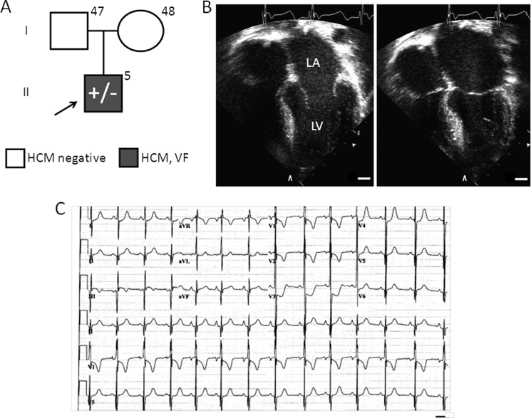
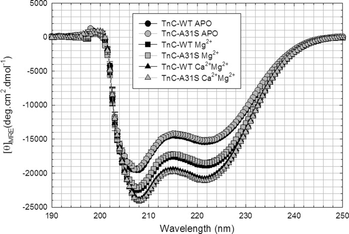
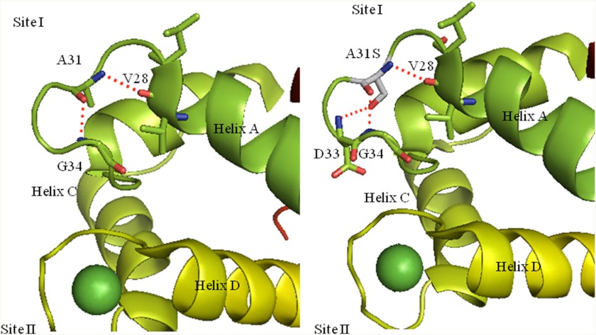
Defined as clinically unexplained hypertrophy of the left ventricle, hypertrophic cardiomyopathy (HCM) is traditionally understood as a disease of the cardiac sarcomere. Mutations in TNNC1-encoded cardiac troponin C (cTnC) are a relatively rare cause of HCM. Here, we report clinical and functional characterization of a novel TNNC1 mutation, A31S, identified in a pediatric HCM proband with multiple episodes of ventricular fibrillation and aborted sudden cardiac death. Diagnosed at age 5, the proband is family history-negative for HCM or sudden cardiac death, suggesting a de novo mutation. TnC-extracted cardiac skinned fibers were reconstituted with the cTnC-A31S mutant, which increased Ca(2+) sensitivity with no effect on the maximal contractile force generation. Reconstituted actomyosin ATPase assays with 50% cTnC-A31S:50% cTnC-WT demonstrated Ca(2+) sensitivity that was intermediate between 100% cTnC-A31S and 100% cTnC-WT, whereas the mutant increased the activation of the actomyosin ATPase without affecting the inhibitory qualities of the ATPase. The secondary structure of the cTnC mutant was evaluated by circular dichroism, which did not indicate global changes in structure. Fluorescence studies demonstrated increased Ca(2+) affinity in isolated cTnC, the troponin complex, thin filament, and to a lesser degree, thin filament with myosin subfragment 1. These results suggest that this mutation has a direct effect on the Ca(2+) sensitivity of the myofilament, which may alter Ca(2+) handling and contribute to the arrhythmogenesis observed in the proband. In summary, we report a novel mutation in the TNNC1 gene that is associated with HCM pathogenesis and may predispose to the pathogenesis of a fatal arrhythmogenic subtype of HCM.


Figures

FIGURE 1.
FIGURE 1.
TNNC1-A31S proband clinical characteristics. A, shown is a pedigree depicting the family of the proband (arrow) who hosts the TNNC1-A31S heterozygous allele as well as his two parents, who are negative for a family history of HCM and sudden cardiac death and are unremarkable upon echocardiographic evaluation. LA, left atrium; LV, left ventricle; VF, ventricular fibrillation; bar, 10 mm. B, shown are representative echocardiographic images demonstrating asymmetric left ventricular hypertrophy and left atrial dilatation during end diastole (left image) and systole (right). C, shown is representative 12-lead electrocardiographic tracing demonstrating voltage criteria for HCM and QT prolongation. Bar, 0.4 s.
FIGURE 2.
FIGURE 2.
Skinned cardiac fiber experiments reconstituted with the HCM cTnC mutant. A, porcine cardiac fibers were reconstituted with mutant (gray circles) and WT cTnC (black circles), and Ca2+ sensitivity of force development was reported as pCa50 values in Table 1; B). Force recovery values (P) obtained after reconstitution with mutant and WT cTnC were compared with the level of force present before extraction of native cTnC (P0) and were reported as % (P/P0). Data are reported as the mean ± S.E. (n = 8–10).
FIGURE 3.
FIGURE 3.
Effect of the HCM cTnC mutant on actin-tropomyosin-troponin-activated ATPase activity measurements. A, activation of actomyosin ATPase by preformed troponin WT and HCM-cTnC mutant complex at increasing ratios is indicated on the abscissa in the presence of Ca2+. B, shown is inhibition of the actomyosin ATPase activity by increasing ratios of preformed troponin WT and HCM-cTnC complex in the absence of Ca2+. For A and B, each point represents an average of six to seven experiments performed in triplicate and is expressed as the mean ± S.E. C, shown is actin-tropomyosin-activated myosin ATPase activity of HCM-cTnC mutant as a function of pCa (each point represents an average of eight experiments, performed in triplicate and expressed as mean ± S.E.). The dark gray circles indicate the presence of 100% cTnC mutant complexes, and light gray circles with the dotted line represent experiments performed with a 50:50 ratio of HCM-cTnC mutant to WT complexes. Black circles represent data obtained with WT alone. The myosin ATPase activity that occurs in the absence of troponin complex is considered 100% ATPase activity. The specific ATPase activity in the absence of troponin complexes was measured as 0.35 mol of Pi × mol of myosin−1 × s−1.
FIGURE 4.
FIGURE 4.
Steady state fluorescence to determine the apparent Ca2+ affinities of IAANS-labeled cTnC A31S mutant. A, isolated cTnC double-labeled with IAANS at Cys-35 and Cys-84 compare Ca2+ titration of A31S (dark gray circles) versus WT (black circles). B, troponin complex with cTnC-IAANS double-labeled at Cys-35 and Cys-84 compare A31S versus WT. C, thin filament with cTnC-IAANS single-labeled at Cys-84 of A31S versus WT is shown. D, thin filament with cTnC-IAANS single-labeled at Cys-84 in the presence of myosin S1 with conditions that favor strong cross-bridge formation (−ATP) is shown. Relative fluorescence values (%) are plotted as a function of Ca2+ concentrations in moles. Data are reported as mean ± S.E. (n = 4–7).
FIGURE 5.
FIGURE 5.
Determination of secondary structural characteristics of HCM mutant and WT-cTnC by circular dichroism. These spectra compare the changes in α-helical content of WT versus cTnC-A31S under different conditions. The graph compares cTnC in the apo, Mg2+-bound, and Mg2+/Ca2+- bound states.
FIGURE 6.
FIGURE 6.
Molecular visualization of the A31S mutation in the crystal structure 1AJ4. A, the H-bonds made by Ala-31 to adjacent residues located in inactive Ca2+ binding site I are shown. B, shown are the putative H-bonds made when the A31S substitution is present.

References

    1. Maron B. J., Epstein S. E., Roberts W. C. (1986) Causes of sudden death in competitive athletes. J. Am. Coll. Cardiol. 7, 204–214 - PubMed
    1. Maron B. J., Gardin J. M., Flack J. M., Gidding S. S., Kurosaki T. T., Bild D. E. (1995) Prevalence of hypertrophic cardiomyopathy in a general population of young adults. Echocardiographic analysis of 4111 subjects in the CARDIA study. Coronary artery risk development in (young) adults. Circulation 92, 785–789 - PubMed
    1. Maron B. J., Roberts W. C., McAllister H. A., Rosing D. R., Epstein S. E. (1980) Sudden death in young athletes. Circulation 62, 218–229 - PubMed
    1. Bos J. M., Towbin J. A., Ackerman M. J. (2009) Diagnostic, prognostic, and therapeutic implications of genetic testing for hypertrophic cardiomyopathy. J. Am. Coll. Cardiol. 54, 201–211 - PubMed
    1. Landstrom A. P., Ackerman M. J. (2010) Mutation type is not clinically useful in predicting prognosis in hypertrophic cardiomyopathy. Circulation 122, 2441–2449 - PMC - PubMed

Publication types

MeSH terms