. 2012 Sep 14;287(38):32096-102.
doi: 10.1074/jbc.M112.355644. Epub 2012 Jul 19.

Structural insights into the anti-methicillin-resistant Staphylococcus aureus (MRSA) activity of ceftobiprole

Affiliations

Structural insights into the anti-methicillin-resistant Staphylococcus aureus (MRSA) activity of ceftobiprole

Andrew L Lovering et al. J Biol Chem. .

Abstract

Methicillin-resistant Staphylococcus aureus (MRSA) is an antibiotic-resistant strain of S. aureus afflicting hospitals and communities worldwide. Of greatest concern is its development of resistance to current last-line-of-defense antibiotics; new therapeutics are urgently needed to combat this pathogen. Ceftobiprole is a recently developed, latest generation cephalosporin and has been the first to show activity against MRSA by inhibiting essential peptidoglycan transpeptidases, including the β-lactam resistance determinant PBP2a, from MRSA. Here we present the structure of the complex of ceftobiprole bound to PBP2a. This structure provides the first look at the molecular details of an effective β-lactam-resistant PBP interaction, leading to new insights into the mechanism of ceftobiprole efficacy against MRSA.


Figures

FIGURE 1.
FIGURE 1.
Chemical structures of MRSA-active and -inactive cephalosporins along with electron density maps and models of ceftobiprole covalently bound to Ser-403 at the PBP2a active site. a, general chemical structure of the cephalosporin antibiotics, indicating the positions of variable R1 and R2 groups and chemical structures of ceftobiprole, nitrocefin, and methicillin. b, final model and 2FoF c map contoured at 1σ. The ceftobiprole R1 group is shown in both of its proposed conformations A and B, with all atoms equivalent up to C7[C2] (indicated by an arrow in panel a). c, results of test refinement with ceftobiprole in the A conformation, with 2FoF c (blue, 1σ) and FoF c (green, 3σ) maps shown. The FoF c difference density is continuous with the modeled and unmodeled 2FoF c electron density, suggesting either multiple conformations or chemical modification of the terminal heterocycle of R1. With no evidence for the latter, and precedent for multiple R1 orientations among other transpeptidase structures, the modeling of two conformations was chosen. d, as in c, with ceftobiprole modeled and test refined in the B conformation. Ceftobiprole is colored by CMYK convention (C, yellow; N, blue; O, red; S, green), and σ represents the root mean square electron density in the crystallographic unit cell.
FIGURE 2.
FIGURE 2.
Structural comparisons demonstrating PBP2a structural rearrangements upon binding of ceftobiprole and the importance of serine nucleophile exposure. a, PBP2a apo structure (orange) compared with PBP2a in complex with ceftobiprole (enzyme in green, ceftobiprole atomic bonds in black, and electron density contoured at 1σ in gray). Secondary structure is shown as transparent ribbons, with backbone and side chain atoms overlaid in solid color. Upon binding, structural rearrangements must take place to accommodate ceftobiprole. These include a shift (toward the left in the figure) of the β-sheet, most notably at strand β3, as well as a clockwise twist (relative to the direction of β3) of the residue backbone and side chain atoms. The arrow diagram illustrates these rearrangements, and the inset depicts the whole PBP2a molecule with the active site indicated by the inhibitor (black). PBP2a domains are shown as follows: N-terminal extension (NTE), non-penicillin-binding domain (NPB), and transpeptidase domain (TP). b, methicillin-susceptible endogenous PBP2 (blue) superimposed on the resistant PBP2a (orange). The Oγ atom of the PBP2 serine nucleophile (Ser-398) is 2.0 Å distant from that of PBP2a Ser-403 and so is far more exposed at the opening of the active site cleft, presumably contributing to its greater availability for acylation in vivo.
FIGURE 3.
FIGURE 3.
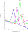
Differential scanning calorimetry traces for PBP2a and acyl-enzyme complexes formed with β-lactams. A (blue), heat capacity change in the presence of 1 mm imipenem. B (magenta), heat capacity change in the presence of 100 μm benzylpenicillin. C (green), heat capacity change observed for native protein. D (red), heat capacity change in the presence of 10 μm ceftobiprole. Each β-lactam was also present in the reference cell, and traces in the absence of protein showed only a steady decline over the temperature range studied (data not shown).

References

    1. Neu H. C. (1992) The crisis in antibiotic resistance. Science 257, 1064–1073 - PubMed
    1. Lyon B. R., Skurray R. (1987) Antimicrobial resistance of Staphylococcus aureus: genetic basis. Microbiol. Rev. 51, 88–134 - PMC - PubMed
    1. Barrett F. F., McGehee R. F., Jr., Finland M. (1968) Methicillin-resistant Staphylococcus aureus at Boston City Hospital: bacteriologic and epidemiologic observations. New Engl. J. Med. 279, 441–448 - PubMed
    1. Doshi R. K., Patel G., Mackay R., Wallach F. (2009) Healthcare-associated infections: epidemiology, prevention, and therapy. Mt. Sinai J. Med. 76, 84–94 - PubMed
    1. Sievert D. M., Rudrik J. T., Patel J. B., McDonald L. C., Wilkins M. J., Hageman J. C. (2008) Vancomycin-resistant Staphylococcus aureus in the United States, 2002–2006. Clin. Infect. Dis. 46, 668–674 - PubMed

Publication types

MeSH terms

Associated data