. 2012 Jul 18;32(29):9785-95.
doi: 10.1523/JNEUROSCI.0482-12.2012.

Identification of the transmitter and receptor mechanisms responsible for REM sleep paralysis

Affiliations

Identification of the transmitter and receptor mechanisms responsible for REM sleep paralysis

Patricia L Brooks et al. J Neurosci. .

Abstract

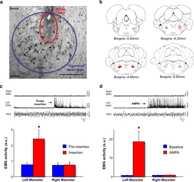
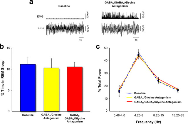
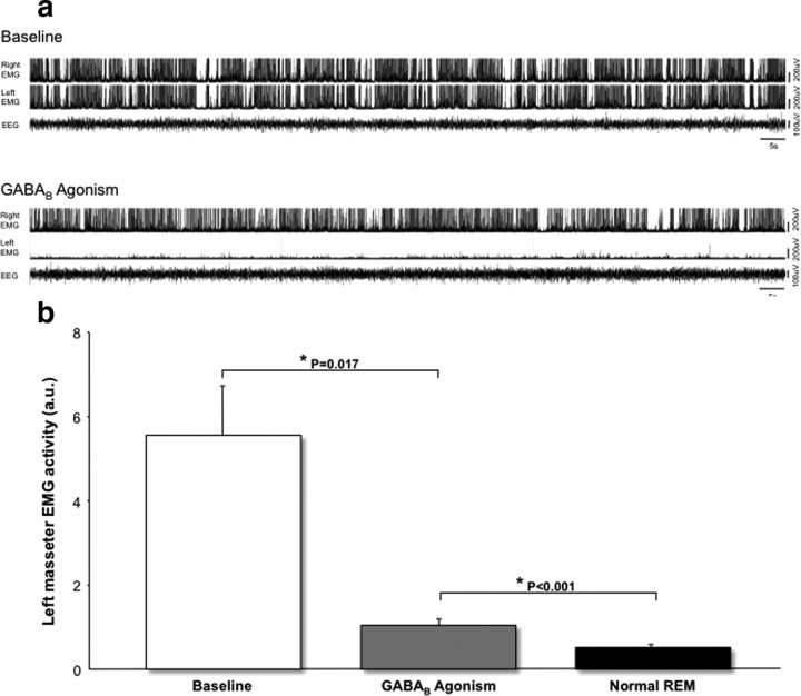
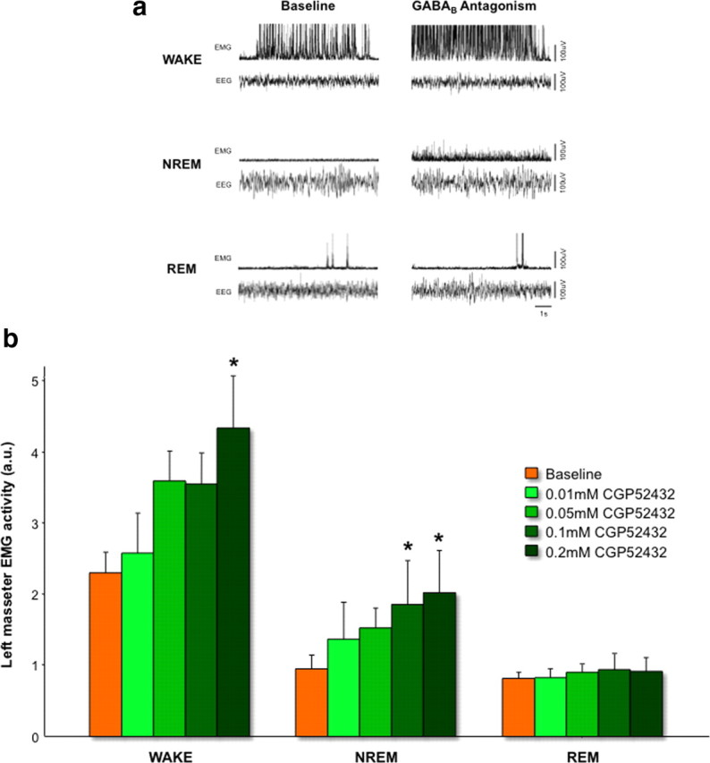
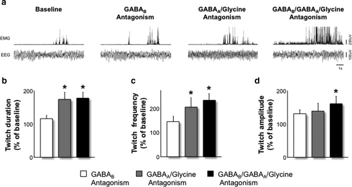
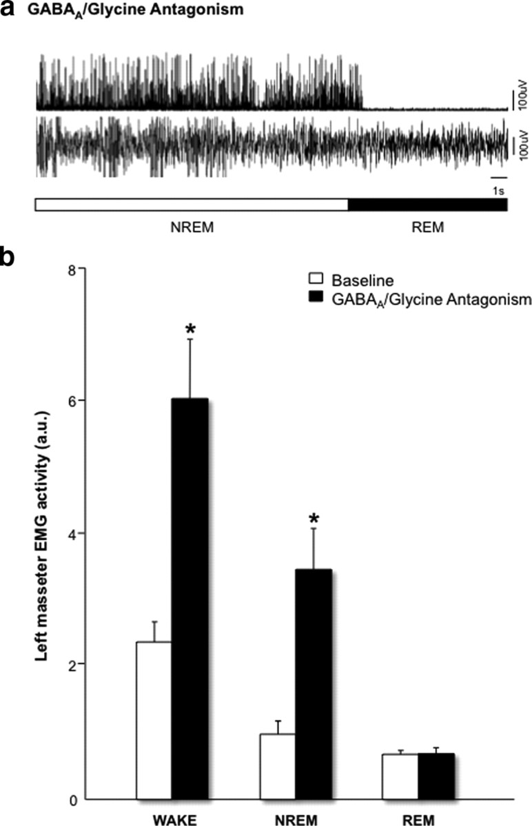
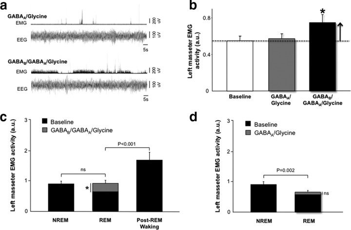
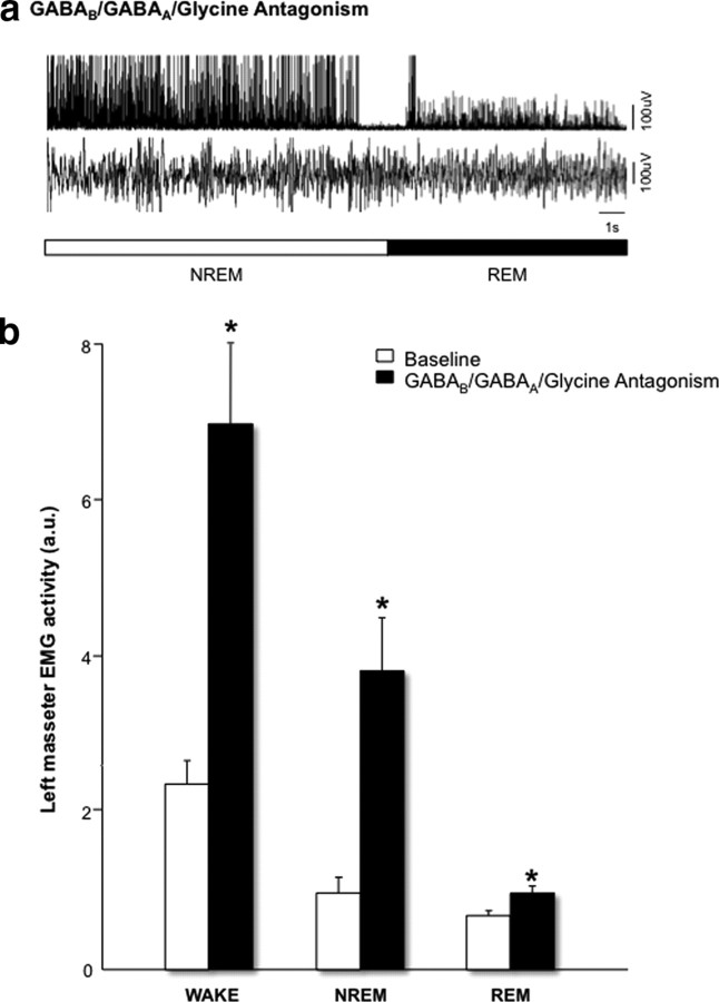
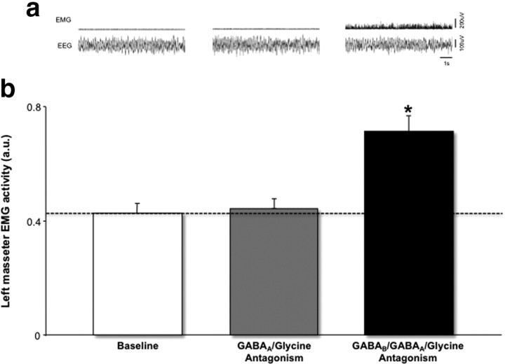
During REM sleep the CNS is intensely active, but the skeletal motor system is paradoxically forced into a state of muscle paralysis. The mechanisms that trigger REM sleep paralysis are a matter of intense debate. Two competing theories argue that it is caused by either active inhibition or reduced excitation of somatic motoneuron activity. Here, we identify the transmitter and receptor mechanisms that function to silence skeletal muscles during REM sleep. We used behavioral, electrophysiological, receptor pharmacology and neuroanatomical approaches to determine how trigeminal motoneurons and masseter muscles are switched off during REM sleep in rats. We show that a powerful GABA and glycine drive triggers REM paralysis by switching off motoneuron activity. This drive inhibits motoneurons by targeting both metabotropic GABA(B) and ionotropic GABA(A)/glycine receptors. REM paralysis is only reversed when motoneurons are cut off from GABA(B), GABA(A) and glycine receptor-mediated inhibition. Neither metabotropic nor ionotropic receptor mechanisms alone are sufficient for generating REM paralysis. These results demonstrate that multiple receptor mechanisms trigger REM sleep paralysis. Breakdown in normal REM inhibition may underlie common sleep motor pathologies such as REM sleep behavior disorder.


Figures

Figure 1.
Figure 1.
Drug interventions preferentially target trigeminal motoneurons. a, A histological example showing the tip of a probe tract (red circle) in the trigeminal motor pool (blue circle). Scale bar, 500 μm. b, Locations of the 31 probe tracts in the trigeminal motor pool plotted on standardized brain maps. Red dots represent probe tip locations in the trigeminal motor pool. Although probes were only placed in the left motor pool, we plotted probe locations in both left and right motor pools in this figure for the sake of visual clarity. c, EEG and EMG traces (top) and group data (bottom) showing that inserting a probe into the left trigeminal motor pool only increases left masseter muscle activity (left EMG), right masseter (right EMG) activity is unaffected. d, EEG and EMG traces (top) and group data (bottom) showing that AMPA perfusion at the left motor pool only increases left masseter muscle activity. *p < 0.05. All values are mean ± SEM.
Figure 2.
Figure 2.
Drug manipulations do not affect REM-generating circuits. a, REM sleep paralysis is abolished by preventing GABA and glycine receptor-mediated inhibition of trigeminal motoneurons. EEG and EMG traces (i.e., masseter muscle) showing that blockade of GABAB/GABAA/glycine receptors on trigeminal motoneurons reversed and prevented masseter REM sleep paralysis. Neither REM sleep amounts (b) nor REM sleep EEG spectral power (c) were affected when ionotropic GABAA/glycine and metabotropic GABAB receptors were antagonized at the trigeminal motor pool. Perfusion of either 0.1 mm bicuculline/strychnine or 0.2 mm CGP52432 and 0.1 mm strychnine/bicuculline at the trigeminal nucleus had no affects on REM sleep amounts or EEG power, indicating that applied drugs did not spread to and influence REM-regulating circuits in the nearby sublaterodorsal nucleus. However, metabotropic GABAB and ionotropic GABAA/glycine receptor antagonism on trigeminal motoneurons had profound affects on masseter tone during REM sleep (a). All values are mean ± SEM.
Figure 3.
Figure 3.
GABAB receptor activation on trigeminal motoneurons reduces masseter tone, but does not trigger muscle paralysis. a, EEG and EMG traces showing that GABAB receptor agonism by baclofen (0.5 mm) perfusion at the left trigeminal motor pool markedly reduces (compared with baseline) left masseter tone (left EMG) during waking. Right masseter muscle tone is unaffected. b, Group data (n = 5) showing that compared with baseline baclofen-induced activation of GABAB receptors on trigeminal motoneurons reduces waking masseter tone. However, this intervention does not reduce waking masseter tone to normal REM sleep levels. All values are mean ± SEM.
Figure 4.
Figure 4.
GABAB receptor antagonism on trigeminal motoneurons does not prevent REM paralysis. a, EEG and EMG traces showing that GABAB receptor blockade by CGP52432 perfusion (0.2 mm) at the trigeminal motor pool causes robust increases in masseter activity during both waking and NREM sleep, but it does not affect levels of masseter tone during REM sleep. b, Group data (n = 7) showing that CGP52432 perfusion (0.01–0.2 mm) heightens masseter EMG activity during waking and NREM sleep, but it does not prevent REM atonia. *p < 0.004. All values are mean ± SEM.
Figure 5.
Figure 5.
Ionotropic GABAA/glycine receptor-mediated inhibition functions to suppress REM muscle twitches. a, EMG and EEG traces illustrating how masseter muscle twitches during REM sleep are affected by antagonism of GABAB (0.2 mm CGP52432), GABAA/glycine (0.1 mm bicuculline/strychnine) and GABAB/GABAA/glycine (0.2 mm CGP52432 and 0.1 mm bicuculline/strychnine) receptors at the trigeminal motor pool. bd, Group data (n = 14) demonstrating how metabotropic, ionotropic, and combined metabotropic/ionotropic receptor blockade on trigeminal motoneurons affects the duration (b), frequency (c) and amplitude (d) of REM muscle twitches. *p < 0.05. All values are mean ± SEM.
Figure 6.
Figure 6.
GABAA and glycine receptor antagonism increases masseter tone during waking and NREM sleep, but it does not prevent REM atonia. a, An EMG and EEG trace showing the abrupt loss of masseter tone on entrance into REM despite continued antagonism of GABAA/glycine receptors. b, Group data (n = 14) showing that bicuculline and strychnine perfusion (0.1 mm for each) onto trigeminal motoneurons increases masseter EMG activity during both waking and NREM sleep. However, this same intervention has no affect on basal levels of muscle tone during REM sleep. *p < 0.001. All values are mean ± SEM.
Figure 7.
Figure 7.
Activation of both metabotropic GABAB and ionotropic GABAA/glycine receptors is required for REM sleep paralysis. a, EMG and EEG traces illustrating that masseter REM atonia remains intact when only GABAA/glycine receptors are antagonized, but prevented and overridden when both GABAB and GABAA/glycine receptors are simultaneously antagonized on trigeminal motoneurons. b, Group data showing that bicuculline and strychnine (0.1 mm) applied onto trigeminal motoneurons during REM sleep cannot prevent REM inhibition (n = 12). However, blockade of both metabotropic GABAB and ionotropic GABAA/glycine receptors by perfusion of CGP52432 (0.2 mm), bicuculline and strychnine (0.1 mm) abolishes masseter REM atonia (n = 13). c, Antagonism of GABAB, GABAA and glycine receptors at the trigeminal motor pool elevates REM masseter tone to baseline NREM sleep levels. However, despite continued receptor antagonism REM masseter tone remains below normal (i.e., baseline) waking levels that occur immediately after REM episodes. This finding indicates that loss of motoneuron excitation helps to reinforce REM muscle paralysis. d, REM paralysis is still triggered even after antagonism of both GABAA/glycine receptors. This graph shows the normal drop in masseter tone when NREM sleep is exited and REM sleep is entered despite continued ionotropic receptor blockade. *p < 0.004. All values are mean ± SEM.
Figure 8.
Figure 8.
Antagonism of GABAB, GABAA and glycine increases basal masseter tone during waking, NREM and REM sleep. a, An EMG and EEG trace showing that REM masseter atonia is not triggered on entrance into REM despite when both metabotropic and ionotropic receptors are antagonized. This is in marked contrast to the complete loss of masseter tone that occurs on entrance into REM sleep when only GABAA/glycine receptors are blocked. b, Group data (n = 13) showing that CGP52432 (0.2 mm) and bicuculline/strychnine perfusion (0.1 mm for each) at the trigeminal motor pool significantly increases basal levels of masseter EMG activity not only during waking and NREM sleep, but also during REM sleep. *p < 0.001. All values are mean ± SEM.
Figure 9.
Figure 9.
REM atonia is triggered by activation of both metabotropic GABAB and iontotropic GABAA/glycine receptors on trigeminal motoneurons. a, EMG and EEG traces showing that REM atonia is reversed when GABAB, GABAA and glycine receptors are antagonized at the trigeminal motor pool; however, atonia remains intact when only GABAA and glycine receptors are blocked. b, Group data showing that even high concentrations of bicuculline and strychnine (0.3 mm) applied onto trigeminal motoneurons during REM sleep cannot prevent REM inhibition (n = 12). However, blockade of both metabotropic GABAB and ionotropic GABAA/glycine receptors by microinjection of CGP52432 (0.6 mm), bicuculline and strychnine (0.2 mm) abolishes masseter REM atonia (n = 13). *p < 0.003. All values are mean ± SEM.

References

    1. Araki T, Yamano M, Murakami T, Wanaka A, Betz H, Tohyama M. Localization of glycine receptors in the rat central nervous system: an immunocytochemical analysis using monoclonal antibody. Neuroscience. 1988;25:613–624. - PubMed
    1. Balasubramanian S, Teissére JA, Raju DV, Hall RA. Hetero-oligomerization between GABAA and GABAB receptors regulates GABAB receptor trafficking. J Biol Chem. 2004;279:18840–18850. - PubMed
    1. Barilà B, Cupello A, Robello M. GABA(B) receptor activation protects GABA(A) receptor from cyclic AMP-dependent down-regulation in rat cerebellar granule cells. Neuroscience. 1999;93:1077–1082. - PubMed
    1. Boissard R, Gervasoni D, Schmidt MH, Barbagli B, Fort P, Luppi PH. The rat ponto-medullary network responsible for paradoxical sleep onset and maintenance: a combined microinjection and functional neuroanatomical study. Eur J Neurosci. 2002;16:1959–1973. - PubMed
    1. Boissard R, Fort P, Gervasoni D, Barbagli B, Luppi PH. Localization of the GABAergic and non-GABAergic neurons projecting to the sublaterodorsal nucleus and potentially gating paradoxical sleep onset. Eur J Neurosci. 2003;18:1627–1639. - PubMed

Publication types

MeSH terms

LinkOut - more resources