. 2012:18:1755-62.
Epub 2012 Jun 27.

Extremely varied phenotypes in granular corneal dystrophy type 2 heterozygotes

Affiliations

Extremely varied phenotypes in granular corneal dystrophy type 2 heterozygotes

Kyung Eun Han et al. Mol Vis. 2012.

Abstract

Purpose: To investigate the phenotypic variability of patients bearing the heterozygous R124H mutation in the TGFBI (transforming growth factor-beta-induced) gene that causes granular corneal dystrophy type 2 (GCD2).

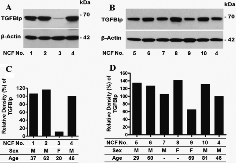
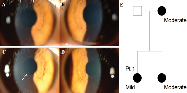
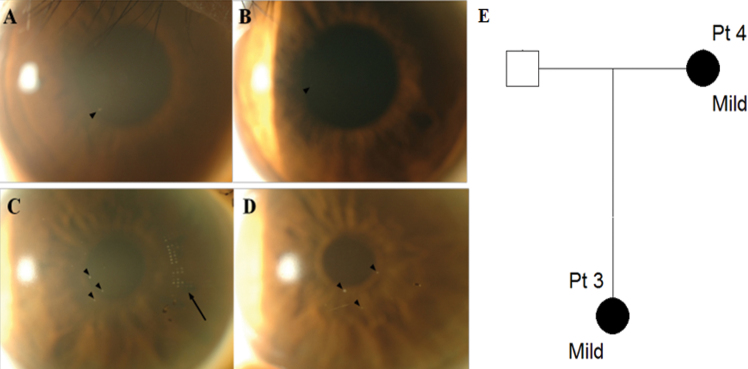
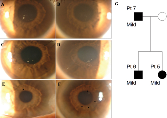
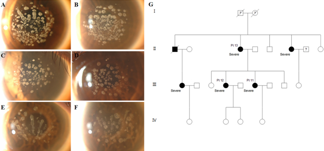
Methods: We describe the phenotypic range of GCD2 heterozygotes for the common R124H mutation in TGFBI; seven with an extremely mild phenotype and six with an extremely severe phenotype. Detailed slit-lamp photographs of these patients were generated. All patients had no history of ocular surgery and were diagnosed as being heterozygous for GCD2 by DNA analysis from peripheral blood. Expression levels of transforming growth factor-beta-induced protein (TGFBIp) were compared among cultured corneal fibroblasts from ten normal donors.

Results: We report profound differences in the severity of the phenotype across our case series. Two patients with a mild phenotype were diagnosed as unaffected at presentation; however follow-up examinations revealed granular deposits. Importantly, we also observed familial clustering of phenotypic variance; five patients from two families with a mild phenotype showed a similarly mild phenotype within family members. Similarly, six patients from two families with severe phenotypes showed corneal deposits with similar patterns and severity within each distinct family, but distinct patterns between families. TGFBIp expressions from different donor derived cultured corneal fibroblasts were different between one another.

Conclusions: GCD2 heterozygotes have extremely varied phenotypes between individual patients. However phenotypes were broadly consistent within families, suggesting that the observed variable expressivity might be regulated by other genetic factors that could influence the abundance of TGFBIp or the function of the pathway. From a clinical perspective, our data also highlighted that genetic analysis and meticulous slit-lamp examination in both eyes at multiple time intervals is necessary.


Figures

Figure 1
Figure 1
Expression of transforming growth factor-beta-induced-gene protein (TGFBIp) in ten different normal corneal fibroblasts (NCF). A, B: western blots show evidence of differential expression of TGFBIp in cultured NCF-1 (37 year-old man), NCF-2 (62 year-old man), NCF-3 (20 year-old woman), NCF-4 (46 year-old man), NCF-5 (29 year-old man), NCF-6 (60 year-old man), NCF-7 (man with unknown-age), NCF-8 (woman with unknown-age), NCF-9 (69 year-old woman), NCF-10 (81 year-old man) from normal donor corneas. In the second experiment, NCF-4 was reloaded for a comparison of the two separate experiments. C, D: Relative density of TGFBIp expression of A and B, respectively. Relative density of NCF-4 was adjusted to 100% in both C and D for a comparison of the densities of normal corneal fibroblasts in two separate experiments. Blots containing 50 μg total protein from normal corneal fibroblasts were incubated with anti-TGFBIp antibody. TGFBIp expression was assayed using anti-TGFBIp polyclonal goat antibodies. β-actin has been used as a control for equal loading. Numbers on the right correspond to the molecular weight markers in kilodaltons (kDa).
Figure 2
Figure 2
Slit-lamp photographs and pedigree of Patient 1 with mild phenotype, aged 23 years. A, C: Right eye, B, D: Left eye. A, B: At presentation, no corneal deposits were observed in both eyes. C, D: After six months, one faint corneal deposit was observed in the right eye (white arrow), but there was still no corneal deposit in the left eye. E: Pedigree of Patient 1. Circles represent women, squares represent men. The filled symbols indicate affected individuals.
Figure 3
Figure 3
Slit-lamp photographs and pedigree of mild family I. A, B: Patient 3, aged 22; C, D: Patient 4, aged 47. A, C: Right eye, B, D: Left eye. A: Granular opacity was observed around pupil margin at 7 o’clock in the anterior stroma in the right eye (black arrowhead). B: Nearly non-detectable corneal deposit was observed in the left eye. C, D: A few dot-like granular deposits (black arrowheads) were observed centrally in the anterior stroma in both eyes. The black arrow in the right eye indicates the mark of a soft contact lens. E: Pedigree of mild family I. Circles represent women, squares represent men. The filled symbols indicate affected individuals.
Figure 4
Figure 4
Slit-lamp photographs and pedigree of mild family II. A, B: Patient 5, aged 33; C, D: Patient 6, aged 34; E, F: Patient 7, aged 62. A, C, E: Right eye, B, D, F: Left eye. A: Several granular and linear corneal opacities were observed. B: A few small granular deposits and linear opacities were observed. C, D: Opacities of round and linear shapes were observed centrally in the anterior stroma in both eyes. E: A small granular deposit was observed around the pupillary margin at 12 o’clock (black arrowhead). F: Faint granular opacities at the 1, 4, 5, and 7 o’clock positions were observed (black arrowheads). G: Pedigree of mild family II. Circles represent women, squares represent men. The filled symbols indicate affected individuals.
Figure 5
Figure 5
Slit-lamp photographs and pedigree of severe family I. A, B: Patient 8, aged 35; C, D: Patient 9, aged 38; E, F: Patient 10, aged 59. A, C, E: Right eye, B, D, F: Left eye. Confluent granular deposits, prominent lattice deposits, and anterior haze occupying the visual axis was observed in both eyes of all family members. They showed a more severe phenotype than other patients of a similar age and members within the family showed a similar pattern. G: Pedigree of severe family I. Circles represent women, squares represent men. The filled symbols indicate affected individuals.
Figure 6
Figure 6
Slit-lamp photographs and pedigree of severe family II. A, B: Patient 11, aged 37; C, D: Patient 12, aged 40; E, F: Patient 13, aged 65. A, C, E: Right eye, B, D, F: Left eye. Numerous granular corneal deposits were observed in both eyes of all family members. They showed a more severe phenotype than other patients of a similar age and members within the family showed a similar pattern. G: Pedigree of severe family II. Circles represent women, squares represent men. The filled symbols indicate affected individuals.

References

    1. Holland EJ, Daya SM, Stone EM, Folberg R, Dobler AA, Cameron JD, Doughman DJ. Avellino corneal dystrophy. Clinical manifestations and natural history. Ophthalmology. 1992;99:1564–8. - PubMed
    1. Okada M, Yamamoto S, Inoue Y, Watanabe H, Maeda N, Shimomura Y, Ishii Y, Tano Y. Severe corneal dystrophy phenotype caused by homozygous R124H keratoepithelin mutations. Invest Ophthalmol Vis Sci. 1998;39:1947–53. - PubMed
    1. Moon JW, Kim SW, Kim TI, Cristol SM, Chung ES, Kim EK. Homozygous granular corneal dystrophy type II (Avellino corneal dystrophy): natural history and progression after treatment. Cornea. 2007;26:1095–100. - PubMed
    1. Konishi M, Yamada M, Nakamura Y, Mashima Y. Varied appearance of cornea of patients with corneal dystrophy associated with R124H mutation in the BIGH3 gene. Cornea. 1999;18:424–9. - PubMed
    1. Rosenwasser GO, Sucheski BM, Rosa N, Pastena B, Sebastiani A, Sassani JW, Perry HD. Phenotypic variation in combined granular-lattice (Avellino) corneal dystrophy. Arch Ophthalmol. 1993;111:1546–52. - PubMed

Publication types

MeSH terms

Substances

Supplementary concepts

LinkOut - more resources