. 2012;7(7):e40707.
doi: 10.1371/journal.pone.0040707. Epub 2012 Jul 17.

A fate map of the murine pancreas buds reveals a multipotent ventral foregut organ progenitor

Affiliations

A fate map of the murine pancreas buds reveals a multipotent ventral foregut organ progenitor

Jesse R Angelo et al. PLoS One. 2012.

Abstract

The definitive endoderm is the embryonic germ layer that gives rise to the budding endodermal organs including the thyroid, lung, liver and pancreas as well as the remainder of the gut tube. DiI fate mapping and whole embryo culture were used to determine the endodermal origin of the 9.5 days post coitum (dpc) dorsal and ventral pancreas buds. Our results demonstrate that the progenitors of each bud occupy distinct endodermal territories. Dorsal bud progenitors are located in the medial endoderm overlying somites 2-4 between the 2 and 11 somite stage (SS). The endoderm forming the ventral pancreas bud is found in 2 distinct regions. One territory originates from the left and right lateral endoderm caudal to the anterior intestinal portal by the 6 SS and the second domain is derived from the ventral midline of the endoderm lip (VMEL). Unlike the laterally located ventral foregut progenitors, the VMEL population harbors a multipotent progenitor that contributes to the thyroid bud, the rostral cap of the liver bud, ventral midline of the liver bud and the midline of the ventral pancreas bud in a temporally restricted manner. This data suggests that the midline of the 9.5 dpc thyroid, liver and ventral pancreas buds originates from the same progenitor population, demonstrating a developmental link between all three ventral foregut buds. Taken together, these data define the location of the dorsal and ventral pancreas progenitors in the prespecified endodermal sheet and should lead to insights into the inductive events required for pancreas specification.


Conflict of interest statement

Competing Interests: The authors have declared no competing interests exist.

Figures

Figure 1
Figure 1. Fate map of the 3–11 SS caudal foregut endoderm.
A–C. Fate maps summarizing the results obtained from DiI labeling the endoderm of individual 3–5 SS (A) 6–8 SS (B) and 9–11 SS (C) embryos and culturing through ∼9.5 dpc. Each shape represents the position and size of the DiI labeled endoderm from a single embryo at the stage indicated by each map. The color indicates which organ bud those labeled cells contributed to at the end of culture. DiI labeled cells that contributed to the dorsal pancreas bud are colored blue, those that contributed to the ventral pancreas bud are yellow, contributors to the liver bud are red while groups of cells which gave rise solely to gut tube are in white. Because multiple embryos were often labeled in similar domains, the color of each shape is transparent and thus darker shades of a color indicate overlapping labeled endoderm that gave rise to the same bud. Labeled groups of cells that gave rise to 2 organ buds are bi-colored and each color placed in the presumptive half that gave rise to the indicated organ. For ease of view, all the data points that gave rise to gut tube alone were placed on top of any colored shapes. A simple compass indicating the embryonic axis is provided. The orientation of the axis is preserved in all fate map cartoons (Figures 2,3,5 and Figure S1).
Figure 2
Figure 2. Identification of the dorsal pancreas progenitors.
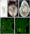
A–B.A cartoon depicting a 9–11 SS embryo labeled over the third somite and slightly lateral on the right side (A) and a bright field/fluorescence merged image of the actual 9 SS embryo revealing the DiI (red) labeled cells (B). C. After culture, the merged image reveals that the resultant 25 SS embryo contained DiI labeled descendants in the dorsal gut (lower red) as well as a smaller patch of visible descendants in the ventral/lateral gut tube (upper red). D–E. Transverse sections as indicated by the dashed lines in (C) revealed the co-localization of DiI (red) in the PDX1-positive (green) dorsal pancreas bud (dp, E) but not in the ventral pancreas bud (vp, D). F–H. A cartoon representing a 5 SS embryo labeled over the third somite pair, as indicated in the cartoon (F) and shown in the actual labeled 5 SS embryo (G) that was cultured to the 20 SS (H). I. Section analysis of the embryo indicated in (H) with PDX1 (green) reveals DiI labeled descendants throughout the dorsal pancreas (arrowheads in I’, boxed region in I is magnified in I’). J. A cartoon of a 5 SS embryo labeled over the left first and second somite, cultured through the 20 SS and processed as above. K–M. Transverse sections through this embryo are aligned in a rostral to caudal manner as indicated by the arrow. DiI labeled descendants (red) are found in the dorsal endoderm rostral to the dorsal pancreas (K) and only slightly overlap with the rostral-most portion of the PDX1-positive dorsal pancreas bud (arrowhead in L). All of the more caudal sections of the dorsal pancreas (green) do not contain DiI labeled cells (M). N. A cartoon depicting a 5 SS embryo labeled in the endoderm over the caudal most fourth and much of the fifth somite. O–Q. Section analysis of the resultant 23 SS embryo, arranged in a rostral to caudal manner as indicated by the arrow, revealed that DiI labeled cells were absent from the rostral regions of the dorsal pancreas bud (green, O) but overlapped slightly with the caudal portion of the dorsal bud (arrowhead, P) and extend into gut tube caudal to the dorsal pancreas bud (red cells in Q). In all sections the arrowheads indicate region of DiI overlap with the dorsal pancreas bud.
Figure 3
Figure 3. Identification of ventral pancreas precursors.
A–C.A 6 SS embryo labeled in the lateral endoderm stretching from the right rostral edge of the AIP to the fourth somite, is depicted in the cartoon (A) and in a merged fluorescent/bright field view of the actual labeled embryo at the onset (red cells in B) and at the end of culture (C, 22 SS). D–E. DiI labeled cells (red, arrowheads) contribute to the liver bud (outlined by the dashed line) and adjacent gut tube (D) as well as in the lateral ventral pancreas (E, arrowheads) denoted by the prominent nuclear PDX1 immunofluorescence (green).
Figure 4
Figure 4. VMEL contribution to multiple foregut organ buds occurs in a temporally restricted manner.
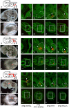
A–B, H–I, O–P. Bright field/fluorescent merged views of embryos labeled with DiI (red) in the VMEL at the onset (A, H, O) and after ∼30 hours of culture (B, I, P). All embryos were sectioned as indicated (dashed lines in B, I and P) and immunofluorescence performed with PDX1 (green) to identify the ventral pancreas bud. Thyroid buds (C, J, Q) and different portions of the liver bud (rostral liver in D, K, R and liver bud in E, L, S) were identified based on morphology. All sections are presented at low magnification (C–F, J–M, Q–T) and a portion of this field (indicated by the boxed area) presented at high magnification (C’–F’, J’–M’, Q’–T’) where the indicated tissue bud has been outlined by a dotted line and arrowheads used to point to DiI labeled cells within the indicated tissue bud. G, N, U. Cartoons summarizing the VMEL contribution (red line) to the thyroid bud (thb), liver bud (lb) and ventral pancreas bud (vpb) for the embryo to the left of the cartoon. A–G. This 5 SS VMEL labeled embryo (red, A) was cultured through the 19 SS (B). Section analysis of this embryo revealed DiI contribution to the thyroid (C, C’), a swath of cells in the rostral liver (D, D’) and a limited number of cells in the midline of the liver (E, E’) and ventral pancreas buds (F, F’) as summarized in G. H–N. The VMEL labeled 5 SS embryo (H) was cultured through the 23 SS (I). Section analysis reveals no VMEL contribution to the thyroid bud (J, J’) but the presence of VMEL descendants in a swath of rostral liver bud (K, K’), in the midline of the liver bud (L, L’) and in the midline of the ventral pancreas bud (M, M’) as summarized (N). O–U. This 8 SS embryo (O) was DiI labeled in the VMEL and cultured through the 28 SS (P). Although labeled VMEL descendants were not found in the thyroid (Q, Q’) or rostral liver buds (R, R’) they were found in the midline of the liver (S, S’) and ventral pancreas buds (T, T’) as summarized (U). v = ventral and d = dorsal gut tube, h = heart.
Figure 5
Figure 5. A summary of the location of foregut organ progenitors during early somite stages.
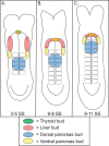
A–C. Cartoons depicting the position of the foregut organ progenitor populations at the 3–5 SS (A), 6–8 SS (B) and 9–11 SS (C) based on the data presented here and elsewhere . The VMEL contribution in the 9–11 SS embryo has not been directly examined and is assumed. The exposed endodermal progenitor populations that are fated to form the 9.5 dpc organ buds are indicated on each map. Blue indicates a dorsal pancreas bud fate, yellow a ventral pancreas bud fate, red a liver bud fate and green a thyroid bud fate. The absence of thyroid progenitors by the 6 SS and the regression of the lateral liver progenitors by the 9 SS indicate that these progenitors have entered the closing foregut. The VMEL population, indicated by the ventral midline oval, produces midline descendants contributing to the thyroid, liver and ventral pancreas buds in a temporally restricted manner.

References

    1. Tam PP, Khoo PL, Lewis SL, Bildsoe H, Wong N, et al. Sequential allocation and global pattern of movement of the definitive endoderm in the mouse embryo during gastrulation. Development. 2007;134:251–260. - PubMed
    1. Cardoso WV, Lu J. Regulation of early lung morphogenesis: questions, facts and controversies. Development. 2006;133:1611–1624. - PubMed
    1. Le Douarin N. An experimental analysis of liver development. Medical Biology. 1975;53:427–455. - PubMed
    1. Gualdi R, Bossard P, Zheng M, Hamada Y, Coleman JR, et al. Hepatic specification of the gut endoderm in vitro: cell signaling and transcriptional control. Genes Dev. 1996;10:1670–1682. - PubMed
    1. Deutsch G, Jung J, Zheng M, Lora J, Zaret KS. A bipotential precursor population for pancreas and liver within the embryonic endoderm. Development. 2001;128:871–881. - PubMed

Publication types