. 2012;7(7):e41105.
doi: 10.1371/journal.pone.0041105. Epub 2012 Jul 17.

Amniotic mesenchymal stem cells enhance wound healing in diabetic NOD/SCID mice through high angiogenic and engraftment capabilities

Affiliations

Amniotic mesenchymal stem cells enhance wound healing in diabetic NOD/SCID mice through high angiogenic and engraftment capabilities

Sung-Whan Kim et al. PLoS One. 2012.

Erratum in

  • PLoS One. 2012;7(10). doi:10.1371/annotation/f6ebe3d3-ef7c-42ce-86fe-d5a661d7f67f

Abstract

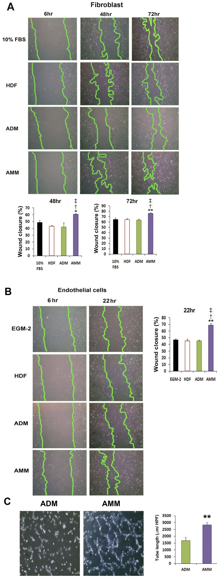
Although human amniotic mesenchymal stem cells (AMMs) have been recognised as a promising stem cell resource, their therapeutic potential for wound healing has not been widely investigated. In this study, we evaluated the therapeutic potential of AMMs using a diabetic mouse wound model. Quantitative real-time PCR and ELISA results revealed that the angiogenic factors, IGF-1, EGF and IL-8 were markedly upregulated in AMMs when compared with adipose-derived mesenchymal stem cells (ADMs) and dermal fibroblasts. In vitro scratch wound assays also showed that AMM-derived conditioned media (CM) significantly accelerated wound closure. Diabetic mice were generated using streptozotocin and wounds were created by skin excision, followed by AMM transplantation. AMM transplantation significantly promoted wound healing and increased re-epithelialization and cellularity. Notably, transplanted AMMs exhibited high engraftment rates and expressed keratinocyte-specific proteins and cytokeratin in the wound area, indicating a direct contribution to cutaneous closure. Taken together, these data suggest that AMMs possess considerable therapeutic potential for chronic wounds through the secretion of angiogenic factors and enhanced engraftment/differentiation capabilities.


Conflict of interest statement

Competing Interests: The authors have declared that no competing interests exist.

Figures

Figure 1
Figure 1. Experimental study design.
Figure 2
Figure 2. Characteristics of AMMs and ADMs.
(A) Microscopic view of AMMs and ADMs (each passage 3). (B) Representative FACS surface markers of AMMs and ADMs. Isotype controls were overlaid in a gray color on each histogram for surface antigen tested.
Figure 3
Figure 3. The expression patterns of angiogenic and and proteins.
(A) qRT-PCR was performed to measure the gene expression levels of HUVECs, HDFs, ADMs, and AMMs. Various cytokines were up-regulated in the AMMs compared with other cell groups. Individual values were normalised to GAPDH. n = 4 per group. ** p<0.01 AMMs vs. ADMs, * p<0.05 AMMs vs. ADMs, p<0.01 AMMs vs. HDFs, p<0.01 AMMs vs. HUVs. Abbreviations: HUV, HUVECs. (B) ELISA for IL-8 and EGF in the cell lysate and supernatant from AMMs, ADMs and HDFs. The amount of IL-8 and IGF-1 were markedly higher in the AMMs than other ADMs and HDFs groups. n = 4 per group. ** p<0.01, * p<0.05, AMMs vs. ADMs, p<0.01 AMMs vs. HDFs. (C) Comparison of the rate of cell apoptosis in response to SD. n = 5 per group. * p<0.05 AMMs vs. ADMs, p<0.01 AMMs vs. HDFs.
Figure 4
Figure 4. Scratch wound and Matrigel tube formation assays.
(A) Representative photograph of fibroblast wound closure after incubation with CM. An in vitro wound healing assay showed that AMM CM strongly improved the fibroblast wound closure compared with the CM of HDF, ADMs, and the control group. (B) Representative photograph of wound closure of endothelial cells after incubation with CM. AMM CM significantly promotes the wound closure of endothelial cells compared with the CM of HUVECs, ADMs, and control. n = 4 per group. ** p<0.01 AMMs vs. ADMs, * p<0.05 AMMs vs. ADMs, p<0.01 AMMs vs. HDFs, p<0.01 AMMs vs. 10% FBS. (C) Representative photograph of Matrigel tube formation of endothelial cells after incubation with CM. n = 6 per group. ** p<0.01 AMMs vs. ADMs.
Figure 5
Figure 5. Effects of AMMs on the in vivo wound closure in mice model and histological analysis of wounds.
(A) Representative photographs of the mouse excisional wound splinting model after transplantations of control vehicle medium (sham), HDFs, ADMs and AMMs at day 0, and 1 and 2 weeks. (B) Wound measurement of each group in STZ-diabetic mice. n = 6 per group. Abbreviations: wk, week(s). (C) Wound histological images by H&E staining. Cross-sectional wound areas were indicated by arrows. (D) Wound histological scores. n = 6 per group. ** p<0.01 AMMs vs. ADMs, * p<0.05 AMMs vs. ADMs, p<0.01 AMMs vs. HDFs, p<0.01 AMMs vs. sham.
Figure 6
Figure 6. Engraftment potential of AMMs.
(A) Representative photographs of localised Dil-labelled ADMs and AMMs. (B) Quantification of engrafted ADMs and AMMs. Dil-labelled cells (red) in the wound area were quantified by histological analysis 4 weeks after cell injection. Dil-labelled cells were injected into the peri-wound areas of NOD/SCID mice. Nuclei were stained with DAPI (blue). n = 5 per group. * p<0.05 AMMs vs. ADMs. (C) Fluorescent in situ hybridization on cell transplanted skin wound tissues. The cells (arrows) exhibited a fluorescent in situ hybridization signal (red) for human chromosome within the nuclei (arrows), suggesting engraftment of injected human cells. (D) Quantification of engrafted ADMs and AMMs by FISH analysis. Wound skin tissues were harvested 4 weeks after cell injection. n = 5 per group. * p<0.05 AMMs vs. ADMs.
Figure 7
Figure 7. Differentiation potential of AMMs.
(A) Representative photographs of cytokeratin-expressing ADMs and AMMs in vivo. Tissue sections from wound area 2 weeks after cell injection were stained with cytokeratin antibody (green). Confocal microscopy analysis showed that cytokeratin (green)-expressing AMMs (red) exist in the epidermis and dermis. Arrows indicate cytokeratin and Dil double-positive cells. (B) Quantification of cytokeratin (green)-positive transplanted (red) ADMs and AMMs. Arrows indicate cytokeratin positive Dil-labeled transplanted cells. ** p<0.01, n = 7.

References

    1. Sumpio BE, Lee T, Blume PA. Vascular evaluation and arterial reconstruction of the diabetic foot. Clin Podiatr Med Surg. 2003;20:689–708. - PubMed
    1. Wu SC, Driver VR, Wrobel JS, Armstrong DG. Foot ulcers in the diabetic patient, prevention and treatment. Vasc Health Risk Manag. 2007;3:65–76. - PMC - PubMed
    1. Moulik PK, Mtonga R, Gill GV. Amputation and mortality in new-onset diabetic foot ulcers stratified by etiology. Diabetes Care. 2003;26:491–494. - PubMed
    1. Campbell WB, Ponette D, Sugiono M. Long-term results following operation for diabetic foot problems: arterial disease confers a poor prognosis. Eur J Vasc Endovasc Surg. 2000;19:174–177. - PubMed
    1. Falanga V. Wound healing and its impairment in the diabetic foot. Lancet. 2005;366:1736–1743. - PubMed

Publication types

MeSH terms