. 2012 Jul 20;150(2):351-65.
doi: 10.1016/j.cell.2012.05.041.

Noncanonical Wnt signaling maintains hematopoietic stem cells in the niche

Affiliations

Noncanonical Wnt signaling maintains hematopoietic stem cells in the niche

Ryohichi Sugimura et al. Cell. .

Abstract

Wnt signaling is involved in self-renewal and maintenance of hematopoietic stem cells (HSCs); however, the particular role of noncanonical Wnt signaling in regulating HSCs in vivo is largely unknown. Here, we show Flamingo (Fmi) and Frizzled (Fz) 8, members of noncanonical Wnt signaling, both express in and functionally maintain quiescent long-term HSCs. Fmi regulates Fz8 distribution at the interface between HSCs and N-cadherin(+) osteoblasts (N-cad(+)OBs that enrich osteoprogenitors) in the niche. We further found that N-cad(+)OBs predominantly express noncanonical Wnt ligands and inhibitors of canonical Wnt signaling under homeostasis. Under stress, noncanonical Wnt signaling is attenuated and canonical Wnt signaling is enhanced in activation of HSCs. Mechanistically, noncanonical Wnt signaling mediated by Fz8 suppresses the Ca(2+)-NFAT- IFNγ pathway, directly or indirectly through the CDC42-CK1α complex and also antagonizes canonical Wnt signaling in HSCs. Taken together, our findings demonstrate that noncanonical Wnt signaling maintains quiescent long-term HSCs through Fmi and Fz8 interaction in the niche.


Figures

Figure 1
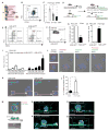
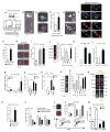
Figure 1. Fmi Regulates Noncanonical Wnt Receptor Fz8 Distribution in Quiescent LT-HSCs
(A and B) Scheme and gating of LT-HSCs, ST-HSCs, and MPPs. (C) qRT-PCR of Fmi in sorted LT-HSCs, ST-HSCs, and MPPs in a setting of triplicates (hereinafter). (D) Scl-tTA-induced H2B-GFP label retention in HSCs. (E) Percentage of LT-HSCs, ST-HSCs, and MPPs in H2B-GFPLSK and H2B-GFPhiLSK. (F) Gating H2B-GFP HSCs. (G–I) qRT-PCR of CD34 (G), Fmi (H), and Fzs (I). (J and K) Immunostaining Fmi and Fz8 in sorted LT-HSCs (J) and H2B-GFP HSCs (K). (L) Percentage of Fmi and Fz8 expressed in sorted H2B-GFP HSCs. (M) Immunostaining Fmi in sorted GFP+LSK cells and OP9 osteoprogenitors. (N–Q) 3D images of Fz8 in LSK on OP9. Scale bar, 5 μM. *p < 0.05. **p < 0.01. ***p < 0.001. Values shown as mean ± SD. See Figure S1.
Figure 2
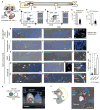
Figure 2. Fmi and Fz8 Colocalize at the Interface between Quiescent LT-HSCs and N-cad+OBs in TBR
(A) HSC and N-cad+OB, Nestin+-MSC, and endothelial cells (CD31+). (B) The sagittal section of Femur. (C and D) Sorted LT-HSCs from TBR (C) and CBR (D) and immunostaining Fmi and Fz8. (E and F) Percentage of LT-HSCs and H2B-GFPhi label-retaining cells (LRCs) in total nuclear cell (TNC) expressing both Fmi and Fz8. (G–V) Location of quiescent HSCs (G and H) or niche components: CD31-GFP (I and J), Nestin-GFP (K and L), N-cad+OB (M–Q). Scale bar, 20 μM. (R–V) Scale bar, 5 μM. (W) Percentage of niche expressing Fmi. (X) 3D image of sorted HSC on OP9. Colocalization of Fmi and Fz8 (yellow arrow) between H2b-GFPhi HSC and OP9 osteoprogenitor. (Y) 3D images of LRC (white, MFI = 122,009 uM2). Colocalization of Fmi and Fz8 (white arrow) at the interface between N-cad+ OB and H2b-GFPhi HSC. *p < 0.05. **p < 0.01. ***p < 0.001. Values shown as mean ± SD. See Figure S2.
Figure 3
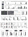
Figure 3. N-cad+OBs Maintain Dominant Noncanonical Wnt Signaling, which Is Attenuated Under Stress
(A) BM section with CD31-GFP, Nestin-GFP, N-cad+OB, and Col2.3-GFP, respectively (shown in green). (B–D) RNA sequencing analysis of sorted niche components for Wnt signaling related genes. See Table S1 for FPKM value. (E and F) Immunostaining TBR. Scale bar, 20 μM. (G) Flow cytometry analysis of N-cad+ OBs percentage in homeostatic (blue) and 5FU-treated (red) conditions. (H and I) Frequency and number of N-cad+OBs in bone cells. (J–L) qRT-PCR analysis of Wnt-related genes. (M and N) Fmi and Fz8 staining in LT-HSCs. (O and P) Measurement of Fmi and Fz8 levels. (Q) Noncanonical Wnt signaling. (R) Ca2+ level in LT-HSCs post-5FU. (S–W) Protein and mRNA levels of LT-HSCs post-5FU: NFAT (S), NFAT targets (T), β-catenin-pS552 (U), TOP-GAL (V), and Axin2 (W). *p < 0.05. **p < 0.01. ***p < 0.001. Values shown as mean ± SD. See Figures S3 and S4.
Figure 4
Figure 4. Fmi and Fz8 Are Required for the Function of LT-HSCs In Vivo
(A and B) Analyses of conventional Fmi (A) or Fz8 (B) knockout mice. Frequency, number, and cell cycle (G0 phase) of HSCs. (C and D) Repopulation analysis of mice receiving transplanted donor cells: 100 LSK (CD45.2) + 2 × 10⁵ BM (CD45.1). Recipients were lethally radiated (CD45.1). Repopulation analysis 16 weeks after injection. (E and F) Frequency and number of donor HSCs 17 weeks posttransplantation. *p < 0.05. **p < 0.01. ***p < 0.001. Values shown as mean ± SD. See Figures S5 and S6.
Figure 5
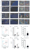
Figure 5. Fmi and Fz8 Maintain Quiescence of LT-HSCs In Vivo
(A–H) Immunostaining of Fz8 (A–D) Fmi (E–H), N-cad, and H2B-GFPhi LRC in TBR and CBR from Wt: Scl-H2B-GFP, Fmi−/−: Scl-H2B-GFP, and Fz8−/−: Scl-H2B-GFP. Scale bar, 20 μM. (I–L) 3D image of protein localization (white arrow). Scale bar, 5 μM. (M and N) Flow cytometry analysis of H2B-GFP intensity in LT-HSCs. (O and P) LRC distribution in TBR and CBR. (Q and R) Frequency of LRC directly contacting N-cad+OBs in TBR. *p < 0.05. **p < 0.01. ***p < 0.001. Values shown as mean ± SD.
Figure 6
Figure 6. Noncanonical Wnt Signaling Mediated by Fz8 Suppresses NFAT-Induced IFNγ Expression and Antagonizes Canonical Wnt Signaling
(A) Ca2+-NFAT and β-catenin pathways. (B and C) LTCC and Fz8 localization in vivo (B) and sorted HSCs (C). (D) Ca2+ level in LT-HSCs in Wt and Fz8−/− mice. (E) NFAT and CK1α localization in LRC of Wt and Fz8−/− mice. (F and G) Fmi, Fz8, and NFAT staining in sorted HSCs. Scale bar, 5 μM. (H and I) LSK from Fmi-shRNA Fz8-shRNA transfected mice were stained with NFAT. (J) NFAT immunostaining of LT-HSCs. (K–O) IFNγ expression in LT-HSCs. (P and Q) β-catenin-pS552 staining (P) and TOP-GAL and Axin2-d2EGFP (Q) staining in LT-HSCs. (R) Axin2 expression in HSCs. (S) Expression of quiescence-related genes. (T) Immunostaining of NFAT. (U and V) Cell-cycle analysis of GFP+LSK cells. (W) Percentage of quiescent HSCs. *p < 0.05. **p < 0.01. ***p < 0.001. Values shown as mean ± SD. See Figure S7.
Figure 7
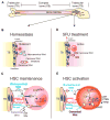
Figure 7. Noncanonical Wnt Signaling Maintains Quiescent LT-HSCs through Fmi and Fz8 Interaction in the Niche
(A) The sagittal section of Femur. (B) N-cad+ OBs maintain a predominant non-canonical Wnt signaling that maintain quiescent HSCs during homeostasis. (C) Fz8 suppresses Ca2+-NFAT nuclear translocation and NFAT-dependent IFNγ expression. (D) Upregulation of canonical Wnt signaling and HSC activation in response to 5FU treatment. (E) Decrease in Fmi-Fz8-mediated noncanonical Wnt signaling resulted from NFAT-induced IFNγ expression and an increase in canonical Wnt signaling, thus together promoting HSC activation.

References

    1. Akashi K, He X, Chen J, Iwasaki H, Niu C, Steenhard B, Zhang J, Haug J, Li L. Transcriptional accessibility for genes of multiple tissues and hematopoietic lineages is hierarchically controlled during early hematopoiesis. Blood. 2003;101:383–389. - PubMed
    1. Arai F, Hirao A, Ohmura M, Sato H, Matsuoka S, Takubo K, Ito K, Koh GY, Suda T. Tie2/angiopoietin-1 signaling regulates hematopoietic stem cell quiescence in the bone marrow niche. Cell. 2004;118:149–161. - PubMed
    1. Baldridge MT, King KY, Boles NC, Weksberg DC, Goodell MA. Quiescent haematopoietic stem cells are activated by IFN-gamma in response to chronic infection. Nature. 2010;465:793–797. - PMC - PubMed
    1. Blanpain C, Fuchs E. Epidermal homeostasis: a balancing act of stem cells in the skin. Nat Rev Mol Cell Biol. 2009;10:207–217. - PMC - PubMed
    1. Butler JM, Nolan DJ, Vertes EL, Varnum-Finney B, Kobayashi H, Hooper AT, Seandel M, Shido K, White IA, Kobayashi M, et al. Endothelial cells are essential for the self-renewal and repopulation of Notch-dependent hematopoietic stem cells. Cell Stem Cell. 2010;6:251–264. - PMC - PubMed

Publication types