. 2012 Jul 19;12(1):97-108.
doi: 10.1016/j.chom.2012.05.016.

An analog of the natural steroidal alkaloid cortistatin A potently suppresses Tat-dependent HIV transcription

Affiliations

An analog of the natural steroidal alkaloid cortistatin A potently suppresses Tat-dependent HIV transcription

Guillaume Mousseau et al. Cell Host Microbe. .

Abstract

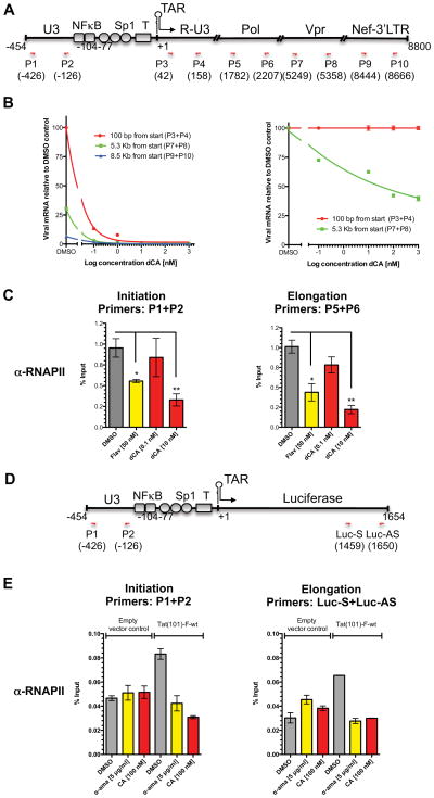
The human immunodeficiency virus type 1 (HIV) Tat protein, a potent activator of HIV gene expression, is essential for integrated viral genome expression and represents a potential antiviral target. Tat binds the 5'-terminal region of HIV mRNA's stem-bulge-loop structure, the transactivation-responsive (TAR) element, to activate transcription. We find that didehydro-Cortistatin A (dCA), an analog of a natural steroidal alkaloid from a marine sponge, inhibits Tat-mediated transactivation of the integrated provirus by binding specifically to the TAR-binding domain of Tat. Working at subnanomolar concentrations, dCA reduces Tat-mediated transcriptional initiation/elongation from the viral promoter to inhibit HIV-1 and HIV-2 replication in acutely and chronically infected cells. Importantly, dCA abrogates spontaneous viral particle release from CD4(+)T cells from virally suppressed subjects on highly active antiretroviral therapy (HAART). Thus, dCA defines a unique class of anti-HIV drugs that may inhibit viral production from stable reservoirs and reduce residual viremia during HAART.


Figures

Figure 1
Figure 1. Structure and activity of dCA on HIV-1 expression
A) Structure of CA and of its analog dCA (B) Activity of dCA on acute replication of HIV-1 at 3 different MOIs. Quantitative CPRG assay. (C) Effect of pre-treating cells with dCA on acute HIV-1 replication. HeLa-CD4 cells were either treated or not with increasing concentrations of dCA. HIV-1 pNL4-3 was added 24 h later to the cells of both experiment sets (MOI≫10) in the presence of testing compound or DMSO control. CPRG assay performed 48 h later. (D) dCA does not block HIV-1 proviral integration into host cells. HeLa-CD4 cells were infected with pNL4-3 (MOI ≫1) in the presence of dCA. Total DNA was extracted 24 h later and integrated provirus was determined by qPCR. (E) Analysis of viral mRNA expression. Total RNA was extracted 3 days after acute infection with pNL4-3 (MOI≫1) in the presence of dCA. First-strand cDNA was quantified by qPCR using primers directed to the env and LTR regions. Results were normalized as copies of viral mRNA per copy of GAPDH mRNA. The arbitrary value of 1 was assigned to the amount of viral mRNA generated in the absence of dCA. RNA samples not reverse transcribed were used as negative control. Error bars represent standard deviations. (F) Viral mRNA expression levels upon dCA treatment of chronically infected cells. HeLa-CD4 cells chronically infected with pNL4-3 were treated with dCA for 10 or 60 days, total RNA was extracted, reverse transcribed and viral cDNA was quantified as in (E). (G) p24 antigen quantification. Viral supernatants recovered from cells described in (B) grown for 60 days, were assayed for their p24 antigen content using a sandwich ELISA kit. Error bars represent standard deviations. (H) Cell viability of HeLa-CD4 cells chronically infected with pNL43 and long term treated with dCA. MTT assay on HeLa-CD4 cells incubated with increasing concentrations of dCA for 12 months. Results are representative of 3 independent experiments. (I) Viral RNA levels upon treatment of CEM-SS cells with dCA and known antiviral drugs. CEM-SS cells chronically infected with pNL4-3 were treated with the indicated compounds for 7 days. Quantification of viral RNA was performed as in (E). Error bars represent standard deviations. Results are representative of 4 independent experiments. See also Figure S1.
Figure 2
Figure 2. dCA binds to Tat and inhibits Tat transactivation of the HIV-1 LTR
(A) dCA prevents transactivation of the HIV-1 promoter by recombinant Tat. HeLa-CD4-LTR-Luc cells were treated with 0.1 μM recombinant Tat protein with increasing concentrations of dCA. Chloroquine 100 μM was added to increase Tat uptake and was added to all points except untreated wells. Luciferase activity was measured 24 h later using the Luciferase Assay System (Promega). Luciferase activity per protein concentration of each sample is shown as relative light units (RLU). HI: Heat Inactivated. (B) HeLa-LTR-Luc cells were transfected with 2 μg of a construct expressing Tat-flag driven by the thymidine kinase (TK) promoter. Twenty-four hours later cells were split and treated or not with dCA at the indicated concentrations. RLU determined 40 h later as in (A). DMSO point set as 100% activation. (C) Schematic diagram of the HIV-1 Tat protein. Depicted above is the amino acid sequence of the wild-type basic domain or a mutated form deficient in binding to the TAR loop. (D) Structure of biotinylated dCA ( Bio-dCA) (E) dCA binds to TAR but not to TAR non-binding mutant of Tat. HEK293T cells were transfected with flag-tagged Tat 101 a.a. (Tat-F-101-wt), a shorter Tat version with 86 a.a. (Ta(86)-F-wt), Tat 86 a.a. mutated in the basic domain (Tat(86)-F-BRM), flag-tagged CDK11 (CDK11-F), flag-tagged 9G8 (9G8-F) and empty vector control. Protein extracts recovered 40 h later were incubated with DynabeadsMyOne Streptavidin T1 coated with either Biotin or Bio-dCA. Pulled-down proteins were revealed by western bloting with the indicated antibodies. (F) dCA interacts with purified recombinant Tat-wt protein. Recombinant Tat-wt protein was incubated with Bio-dCA streptavidin-coated beads in the presence or absence of an excess of non-biotinylated dCA (represented as molar equivalent [eq.] of Bio-dCA) and Raltegravir used as a negative control competitor. Recombinant GST-9G8 and Tat-BRM were used as negative protein-binding control. Pulled-down proteins were revealed by western blot with anti-Tat and anti-GST antibodies. See also Figure S2.
Figure 3
Figure 3. dCA alters Tat-Flag cellular localization
Confocal microscopy analysis of the sub-cellular localization of transfected Tat(86)-F-wt and Tat(86)-F-BRM and where indicated treated (24 h) or not with dCA. Flag epitope-tagged Tat was recognized with anti-flag and AlexaFluor 568 anti-IgG. Transfections were performed in HeLa-CD4 cells. Magnification 300X. See also Figure S3.
Figure 4
Figure 4. RNAPII elongation from viral promoter is inhibited by dCA
(A) Schematic representation of the HIV genome and localization of primers. (B) HIV-1 elongation from HIV-1 promoter measured by qPCR. Left panel: Total RNA was recovered from chronically infected HeLa cells treated with increasing amounts of dCA for 60 days and viral cDNA was measured by relative RT-qPCR using the indicated primers located either at 100 bp, 5.3 kb or 8.5 kb downstream from the transcript start site. All messages normalized to GAPDH mRNA. The amounts of viral mRNA generated in the absence of compound, measured at 100 bp from start site, arbitrarily set at 100%. Right panel: Plotting of data obtained in left panel, setting each data point obtained with the 100 bp primer set at 100% and comparing to results obtained with 5.3 kb primer set. Error bars represent standard deviations of the RT-qPCR assay. Results are representative of 3 independent experiments. (C) ChIP assay of the HIV promoter. HeLa-CD4 cells chronically infected with pNL4-3 were treated with dCA for 4 days and flavopiridol (Flav) for 6–8 h followed by RNAPII ChIP. DNA was measured by qPCR using the indicated set of primers. Results represented as percentages of input. Error bars, ± S.E.M. from 3 independent experiments. *P < 0.05; **P < 0.01 (unpaired t-test). Background was an average of 0.1 ± 0.02 and 0.02 ± 0.01 (S.E.M.) for initiation and elongation respectively. (D) Schematic representation of the LTR-Luciferase transgene and localization of primers. (E) ChIP assay of the LTR-Luciferase. HeLa-CD4-LTR-Luc cells were transfected with pGL4.74-Tat(101)-F-wt or empty vector control and treated with dCA (100 nM) or α-amanitin (α-ama) (5 μg/ml) for 48 h followed by protein-DNA crosslinking and ChIP as in (A) the indicated set of primers. The ChIP background was an average of 0.008± 0.0006 (S.E.M.). Error bars from qPCR (n=3), ± S.E.M. This is representative of 2 independent experiments. See also Figure S4.
Figure 5
Figure 5. Effect of dCA on acute replication of HIV-1 compared with known retroviral inhibitors
(A–D) HeLa-CD4 cells were infected with HIV-1 pNL4-3 in the presence of the indicated compounds or DMSO. Forty hours post infection a CPRG assay was performed. Same dCA curve plotted in graphs (A–D). Error bars represent standard deviation. (E) Analysis of viral mRNA expression upon treatment with dCA and retroviral inhibitors. Total RNA extracted 4 days after acute infection with pNL4-3 (MOI≫1) in the presence of drugs. Viral cDNA was quantified by qPCR using primers recognizing the env and LTR regions. Results normalized to copies of GAPDH mRNA, with value of 1 assigned to the DMSO control. Error bars represent standard deviation. (F) Activity of dCA on acute HIV-2 replication. HeLa-CD4 cells infected with HIV-2 ROD/A in the presence of the indicated concentrations of dCA. Antigen p27 in the supernatant measured 5 days post-infection. (G) Activity of dCA on chronic HIV-2 replication. HeLa-CD4 cells chronically infected with ROD/A were treated with the indicated concentrations of dCA and antigen p27 measured 7 days post-treatment. (H–I) Activity of dCA on primary cells. Freshly isolated uninfected human PBMCs were stimulated with PHA for 2 days, washed, and grown from then on in IL-2 alone. Cells were infected with pNL4-3 in the presence of increasing concentrations of dCA, saquinavir and raltegravir. Antigen p24 was measured 5 days post infection. (J) No viral rebound upon termination of dCA treatment. Left: HeLa-CD4-LTR-LacZ cells chronically infected with pNL4-3 were treated with dCA for 60 days and treated with dCA for another 10 days (dCA) or stopped dCA treatment for 10 days (dCA Stop). The viral mRNA copies were quantified by qPCR and normalized with GAPDH mRNA. Viral mRNA output from untreated cells was assigned as 1. RNA extracts were used in the qPCR as negative control for the presence of genomic DNA contamination, 0.1% of the amplification of the cDNA samples is due to genomic DNA. The error represents a covariance of less than 5%. Right: Same as left, however treatment was stopped for 27 days before harvest of the cells.
Figure 6
Figure 6. dCA activity on CD4+T cells from viremic and aviremic HIV-infected subjects
(A) dCA effect on CD4+T cells isolated from viremic subjects. (A) Viral production from CD4+T cells isolated from 8 viremic subjects and grown in IL-2 was measured in the presence or absence of ARVs (RAL+AZT+EFV) supplemented or not with 100 nM dCA (circle) or 1μM dCA (square). Viral production in the supernatant measured by ELISA p24 and normalized to the negative control (Mock). Statistical significance was assessed by a paired t-test. (B) dCA effect on CD4+T cells isolated from aviremic subjects. CD4+T cells isolated from PBMCs from 4 subjects who had been treated with HAART for at least 3 years were cultured for 6 days without IL-2 in the presence of ARVs to block de novo infection. dCA (100 nM) effect on the spontaneous release of HIV particles was assessed by measuring viral RNA in culture supernatants by ultrasensitive RT-qPCR. See also figure S6.

Comment in

References

    1. Alcami J, Lain de Lera T, Folgueira L, Pedraza MA, Jacque JM, Bachelerie F, Noriega AR, Hay RT, Harrich D, Gaynor RB, et al. Absolute dependence on kappa B responsive elements for initiation and Tat-mediated amplification of HIV transcription in blood CD4 T lymphocytes. Embo J. 1995;14:1552–1560. - PMC - PubMed
    1. Aoki S, Watanabe Y, Sanagawa M, Setiawan A, Kotoku N, Kobayashi M. Cortistatins A, B, C, and D, anti-angiogenic steroidal alkaloids, from the marine sponge Corticium simplex. J Am Chem Soc. 2006;128:3148–3149. - PubMed
    1. Aoki S, Watanabe Y, Tanabe D, Arai M, Suna H, Miyamoto K, Tsujibo H, Tsujikawa K, Yamamoto H, Kobayashi M. Structure-activity relationship and biological property of cortistatins, anti-angiogenic spongean steroidal alkaloids. Bioorg Med Chem. 2007;15:6758–6762. - PubMed
    1. Arima N, Kuziel WA, Grdina TA, Greene WC. IL-2-induced signal transduction involves the activation of nuclear NF-kappa B expression. J Immunol. 1992;149:83–91. - PubMed
    1. Aris JP, Blobel G. cDNA cloning and sequencing of human fibrillarin, a conserved nucleolar protein recognized by autoimmune antisera. Proc Natl Acad Sci U S A. 1991;88:931–935. - PMC - PubMed

Publication types

MeSH terms