

Heart repair and regeneration: recent insights from zebrafish studies
- PMID: 22818295
- PMCID: PMC3445789
- DOI: 10.1111/j.1524-475X.2012.00814.x
Heart repair and regeneration: recent insights from zebrafish studies
Abstract
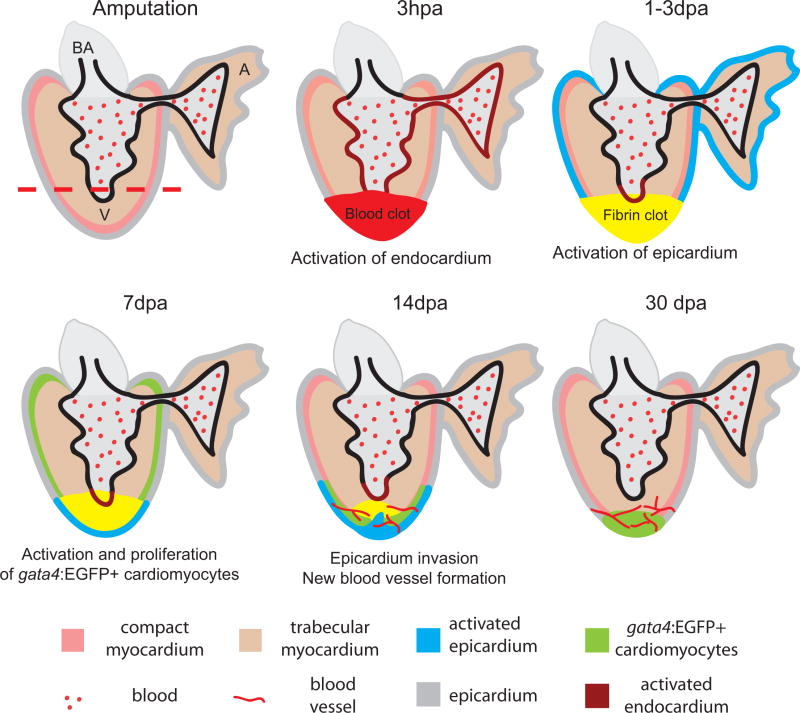
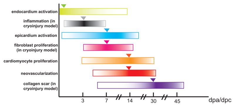
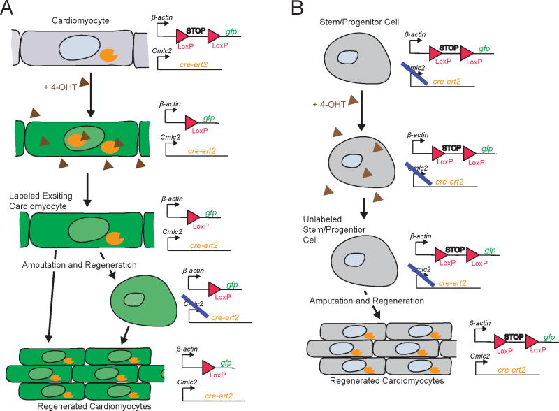
Cardiovascular disease is the leading cause of death in the U.S. and worldwide. Failure to properly repair or regenerate damaged cardiac tissues after myocardial infarction is a major cause of heart failure. In contrast to humans and other mammals, zebrafish hearts regenerate after substantial injury or tissue damage. Here, we review recent progress in studying zebrafish heart regeneration, addressing the molecular and cellular responses in the three tissue layers of the heart: myocardium, epicardium, and endocardium. We also compare different injury models utilized to study zebrafish heart regeneration and discuss the differences in responses to injury between mammalian and zebrafish hearts. By learning how zebrafish hearts regenerate naturally, we can better design therapeutic strategies for repairing human hearts after myocardial infarction.
© 2012 by the Wound Healing Society.
Figures
Similar articles
-
The face of epicardial and endocardial derived cells in zebrafish.Exp Cell Res. 2018 Aug 1;369(1):166-175. doi: 10.1016/j.yexcr.2018.05.022. Epub 2018 May 25. Exp Cell Res. 2018. PMID: 29807022
-
Coronary Revascularization During Heart Regeneration Is Regulated by Epicardial and Endocardial Cues and Forms a Scaffold for Cardiomyocyte Repopulation.Dev Cell. 2019 Nov 18;51(4):503-515.e4. doi: 10.1016/j.devcel.2019.10.019. Dev Cell. 2019. PMID: 31743664 Free PMC article.
-
Retinoic acid production by endocardium and epicardium is an injury response essential for zebrafish heart regeneration.Dev Cell. 2011 Mar 15;20(3):397-404. doi: 10.1016/j.devcel.2011.01.010. Dev Cell. 2011. PMID: 21397850 Free PMC article.
-
Cardiovascular regeneration in non-mammalian model systems: what are the differences between newts and man?Thromb Haemost. 2007 Aug;98(2):311-8. Thromb Haemost. 2007. PMID: 17721612 Review.
-
Cellular and Molecular Mechanism of Cardiac Regeneration: A Comparison of Newts, Zebrafish, and Mammals.Biomolecules. 2020 Aug 19;10(9):1204. doi: 10.3390/biom10091204. Biomolecules. 2020. PMID: 32825069 Free PMC article. Review.
Cited by
-
Improvement of surface ECG recording in adult zebrafish reveals that the value of this model exceeds our expectation.Sci Rep. 2016 Apr 29;6:25073. doi: 10.1038/srep25073. Sci Rep. 2016. PMID: 27125643 Free PMC article.
-
Recent advancements in understanding endogenous heart regeneration-insights from adult zebrafish and neonatal mice.Semin Cell Dev Biol. 2016 Oct;58:34-40. doi: 10.1016/j.semcdb.2016.04.011. Epub 2016 Apr 27. Semin Cell Dev Biol. 2016. PMID: 27132022 Free PMC article. Review.
-
Zebrafish as a Model to Study Vascular Elastic Fibers and Associated Pathologies.Int J Mol Sci. 2022 Feb 14;23(4):2102. doi: 10.3390/ijms23042102. Int J Mol Sci. 2022. PMID: 35216218 Free PMC article. Review.
-
Targeting the hedgehog signaling pathway for cardiac repair and regeneration.Herz. 2017 Nov;42(7):662-668. doi: 10.1007/s00059-016-4500-y. Epub 2016 Nov 22. Herz. 2017. PMID: 27878328 Review. English.
-
Model systems for cardiovascular regenerative biology.Cold Spring Harb Perspect Med. 2013 Apr 1;3(4):a014019. doi: 10.1101/cshperspect.a014019. Cold Spring Harb Perspect Med. 2013. PMID: 23545574 Free PMC article. Review.
References
-
- Lloyd-Jones D, Adams RJ, Brown TM, Carnethon M, Dai S, De Simone G, et al. Executive summary: heart disease and stroke statistics--2010 update: a report from the American Heart Association. Circulation. 2010;121(7):948–54. - PubMed
-
- Bolli P, Chaudhry HW. Molecular physiology of cardiac regeneration. Ann N Y Acad Sci. 2010;1211:113–26. - PubMed
-
- Holmes JW, Borg TK, Covell JW. Structure and mechanics of healing myocardial infarcts. Annu Rev Biomed Eng. 2005;7:223–53. - PubMed
Publication types
MeSH terms
Grants and funding
LinkOut - more resources
Full Text Sources
