Review
. 2012 Aug;36(4):248-59.
doi: 10.1053/j.semperi.2012.04.005.

Effect of processing and storage on red blood cell function in vivo

Affiliations
Review

Effect of processing and storage on red blood cell function in vivo

Allan Doctor et al. Semin Perinatol. 2012 Aug.

Abstract

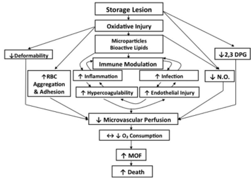
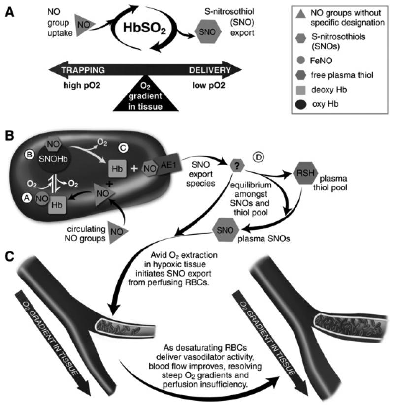
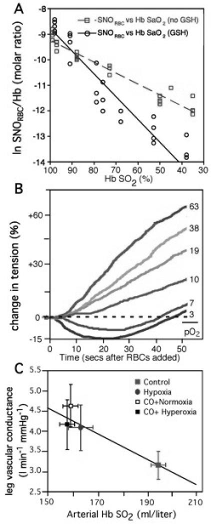
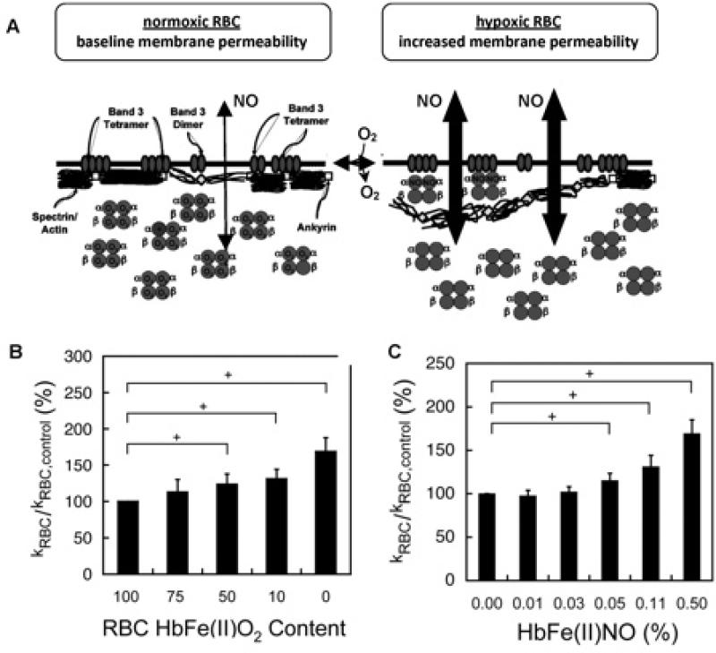
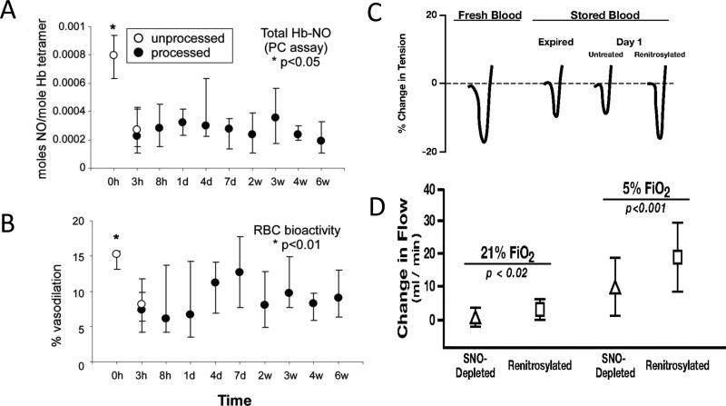
Red blood cell (RBC) transfusion is indicated to improve oxygen delivery to tissue, and for no other purpose. We have come to appreciate that donor RBCs are fundamentally altered during processing and storage in a manner that both impairs oxygen transport efficacy and introduces additional risk by perturbing both immune and coagulation systems. The protean biophysical and physiological changes in RBC function arising from storage are termed the "storage lesion;" many have been understood for some time; for example, we know that the oxygen affinity of stored blood rises during the storage period and that intracellular allosteric regulators, notably 2,3-bisphosphoglyceric acid and ATP, are depleted during storage. Our appreciation of other storage lesion features has emerged with improved understanding of coagulation, immune, and vascular signaling systems. Here, we review key features of the "storage lesion." Additionally, we call particular attention to the newly appreciated role of RBCs in regulating linkage between regional blood flow and regional O(2) consumption by regulating the bioavailability of key vasoactive mediators in plasma, and discuss how processing and storage disturb this key signaling function and impair transfusion efficacy.


Figures

FIGURE 1
FIGURE 1
Interaction between donor RBCs and transfusion recipients as a consequence of receiving RBCs altered by processing and storage. The figure illustrates that the storage lesion impacts overlapping pathways of oxygen delivery, RBC rheology and physiology, as well as immune modulation. MOF = multiple organ failure; NO = nitric oxide.
FIGURE 2
FIGURE 2
RBCs transduce regional O2 gradients in tissue to control NO bioactivity in plasma by trapping or delivering NO groups as a function of Hb O2 saturation. A) In this fashion, circulating NO groups are processed by Hb into the highly vasoactive (thiol-based) NO congener, S-nitrosothiol (SNO). By exporting SNOs as a function of Hb deoxygenation, RBCs precisely dispense vasodilator bioactivity in direct proportion to regional blood flow lack. B) O2 delivery homeostasis requires biochemical coupling of vessel tone to environmental cues that matches perfusion sufficiency to metabolic demand. Because oxy- and deoxy-Hb process NO differently (see text), allosteric transitions in Hb conformation afford context-responsive (O2 - coupled) control of NO bioavailability, thereby linking the sensor and effector arms of this system. Specifically, Hb conformation governs the equilibria among deoxyHbFeNO (A; NO sink), oxySNOHb (B; NO store), and acceptor thiols including the membrane protein SNO-AE1 (C; bioactive NO source). Direct SNO export from RBCs or S-transnitrosylation from RBCs to plasma thiols (D) yields vasoactive SNOs, which influence resistance vessel caliber and close this signaling loop. Thus, RBCs either trap (A) or export (D) NO groups to optimize blood flow. C) NO processing in RBCs (panels A and B) couples vessel tone to tissue PO2; this system subserves hypoxic vasodilation in the arterial periphery and thereby calibrates blood flow to regional tissue hypoxia.
FIGURE 3
FIGURE 3
Hb O2 saturation (Hb SO2%) exerts coordinated governance of RBC SNO content, RBC vasoactivity and human peripheral blood flow. A) Human blood gas measurements of SNO and O2. RBCs with (black) or without (red) added extracellular glutathione (GSH) were deoxygenated under inert gas. The natural logarithm of SNO content in RBCs (SNORBC) is linearly related to Hb O2 saturation. GSH accelerated SNORBC decay, consistent with O2 -linked export of NO groups to extra-erythrocytic thiols. B) RBCs induce graded relaxation of systemic arteries (aortic ring bioassay) that is inversely related to Hb O2 saturation across the physiological range, recapitulating hypoxic vasodilation. Hb O2 saturations spanned from red (oxy, > 90% Hb SO2) to blue (deoxy, < 40% Hb SO2). C) Leg vascular conductance increases as blood O2 content falls (hypoxic vasodilation). Hb O2 saturation and thus arterial blood O2 content were manipulated by CO exposure ± varying FiO2 (fraction of inspired O2; hypoxia versus hyperoxia). Neither vascular conductance nor blood flow correlated with blood PO2 per se.
FIGURE 4
FIGURE 4
Conformation-specific binding between Hb and the RBC membrane protein AE-1 affords O2 responsive control of NO trapping. A) Under oxygenated conditions (normoxic RBC, at left), the RBC membrane constitutes a significant barrier to NO entry via tight association between the sub-membrane cytoskeleton and the cytoplasmic domain of the Band 3 (AE-1) membrane protein (ankyrin successfully competes with oxyHb for binding to AE-1). Upon RBC deoxygenation (hypoxic RBC), oxyHb R-state is converted to deoxyHb T-state, which now successfully competes with ankyrin for the AE-1 cytoplasmic domain (affinity for AE-1 ranks as follows: deoxyHb > ankryn > oxyHb). Proximal apposition of heme to the membrane and diminished cytoskeleton-membrane interaction allows increased NO entry and affords intraerythrocytic Hb greater access to extraerythrocytic NO. Moreover, if deoxyHb encounters high concentrations of NO, “super-T” αFe(II)NOHb may form (in which NO bound to the α subunit disrupts normal heme-globin linkage, locking Hb in the deoxy or T conformation; see text for details). B) Increasing the proportion of intra-erythrocytic T-state Hb (by forming either deoxyHb or αFe(II)NO, both of which are T-state tetramers and bind avidly to AE-1) increases NO consumption by intact RBCs as measured by a competition assay. In this plot, increased consumption in treated vs control RBCs appears as an increased KRBC/KRBCcontrol ratio (y-axis). C) Increased NO uptake by NOpretreated hypoxic RBCs correlates with the formation of αFe(II)NO (i.e. “super-T” Hb).
FIGURE 5
FIGURE 5
O2-dependent variation in SNO–Hb (■) and Hb[FeNO] (□) demonstrate the association between Hb conformation and intramolecular heme → thiol migration of NO groups in RBCs. A - D), Moles NO per mole Hb tetramer in arterial and mixed venous blood from humans breathing either 21% O2 at 1 ATA (absolute atmospheres) (A and D), 21% O2 at 0.56 ATA (equivalent to ~ 12% O2 at 1 ATA) (B) or 100% O2 at 3 ATA (D). Total Hb-bound NO equals the sum of the 2 bars for each condition. These data demonstrate O2 - dependent shuttling of Hb-bound NO groups between heme and Cys-ß93. (E and F), SNO content of blood Hb, presented as the fraction of Hb-NO (% SNO), correlates with Hb O2 saturation (E), but not with pO2 (F).
FIGURE 6
FIGURE 6
PO2 - regulated export of NO groups from RBCs can occur via NO group transfer from SNO-Hb to an extra-erythrocytic thiol reactant. Circulatory transit was simulated for human whole blood in a thin-film tonometer (under 5% CO2, balance N2, pH 7.4) after spiking the sample with the non-native, low-mass thiol, N-acetyl cysteine (NAC, 100 M in plasma). The concentration of S-nitroso-N-acetyl cysteine (SNOAC) that formed in plasma was measured by mass spectrometry, and confirmed by mass labeling with 15N. The conversion of extra-erythrocytic NAC to SNOAC correlated with RBC O2 and SNO content. Serum SNOAC formed as a function of Hb SO2. A) Liquid chromatogram demonstrating co-elution of RBC-generated SNOAC with the 15N-labeled SNOAC standard. B) Mass spectrum demonstrating paired signals from RBC-generated SNOAC and the 15N-labeled SNOAC standard (m/z 194). C) Extra-erythrocytic SNOAC concentration follows oxyHb desaturation (by co-oximetry). D) RBC SNO content (black) decreased in tandem with HbO2 desaturation (dashed blue). Note that, as SNO content in RBCs fell, extra-erythrocytic SNOAC (yellow symbols) accumulated. Note also that SNOAC levels were below the limits of detection when the HbO2 saturation was above 80%.
FIGURE 7
FIGURE 7
Impact of processing and storage upon RBC NO content and vasoactivity (both in isolated vascular rings and in a whole-animal cardiac preparation). In these projects, blood from healthy volunteers was leukofiltered, processed into standard additive solution and stored according to AABB standards. (A) RBC NO content was significantly depressed, (B) as was vasoactivity in a rabbit vascular ring preparation. (C) Representative tracings from a similar project demonstrate the degree of vasorelaxation as percent change in tension in rabbit aortic rings produced by fresh, stored (expired and day 1), or renitrosylated (day 1) RBCs. (D) Hypoxic vasodilation by stored and renitrosylated RBCs in vivo. Shown are changes in canine coronary artery bloodflow(mean ± SD; n7) produced by infusion of SNO-depleted or renitrosylated RBCs. Increases in flow elicited by renitrosylated RBCs were significantly greater than those produced by SNO-depleted RBCs, and the degree of change was greater under hypoxic (5% FiO2) than normoxic (21% FiO2) conditions.

References

    1. Marik PE, Corwin HL. Efficacy of red blood cell transfusion in the critically ill: A systematic review of the literature. Crit Care Med. 2008;36:2667–2674. - PubMed
    1. Spinella PC, Perkins JG, Grathwohl KW, Repine T, Beekley AC, Sebesta J, Jenkins D, Azarow K, Holcomb JB. Risks associated with fresh whole blood and red blood cell transfusions in a combat support hospital. Crit Care Med. 2007;35:2576–2581. - PubMed
    1. Ho J, Sibbald WJ, Chin-Yee IH. Effects of storage on efficacy of red cell transfusion: When is it not safe? Crit Care Med. 2003;31:S687–S697. - PubMed
    1. Marik PE, Sibbald WJ. Effect of stored blood transfusion on oxygen delivery in patients with sepsis. JAMA. 1993;269:3024–3029. - PubMed
    1. Napolitano LM, Corwin HL. Efficacy of blood transfusion in the critically ill: Does age of blood make a difference? Crit Care Med. 2004;32:594–595. - PubMed

MeSH terms