. 2012 Oct;38(10):1716-25.
doi: 10.1016/j.ultrasmedbio.2012.04.015. Epub 2012 Jul 19.

Improved anti-tumor effect of liposomal doxorubicin after targeted blood-brain barrier disruption by MRI-guided focused ultrasound in rat glioma

Affiliations

Improved anti-tumor effect of liposomal doxorubicin after targeted blood-brain barrier disruption by MRI-guided focused ultrasound in rat glioma

Lisa H Treat et al. Ultrasound Med Biol. 2012 Oct.

Abstract

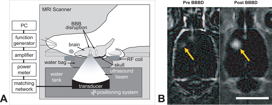
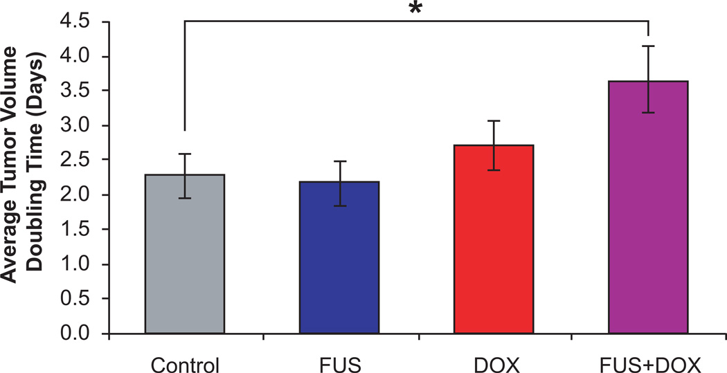
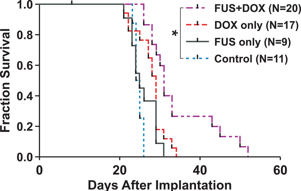
The blood-brain barrier (BBB) inhibits the entry of the majority of chemotherapeutic agents into the brain. Previous studies have illustrated the feasibility of drug delivery across the BBB using focused ultrasound (FUS) and microbubbles. Here, we investigated the effect of FUS-enhanced delivery of doxorubicin on survival in rats with and 9L gliosarcoma cells inoculated in the brain. Each rat received either: (1) no treatment (control; N = 11), (2) FUS only (N = 9), (3) IV liposomal doxorubicin (DOX only; N = 17), or (4) FUS with concurrent IV injections of liposomal doxorubicin (FUS+DOX; N = 20). Post-treatment by magnetic resonance imaging (MRI) showed that FUS+DOX reduced tumor growth compared with DOX only. Further, we observed a modest but significant increase in median survival time after a single treatment FUS+DOX treatment (p = 0.0007), whereas neither DOX nor FUS had any significant impact on survival on its own. These results suggest that combined ultrasound-mediated BBB disruption may significantly increase the antineoplastic efficacy of liposomal doxorubicin in the brain.


Figures

Fig. 1
Fig. 1
A, Experimental set-up for MRI-guided focused ultrasound-induced BBB disruption in rats with implanted 9L gliosarcoma. Pulsed ultrasound was focused in and around the tumor (Frequency: 1.7 MHz, Pressure: 1.2 MPa, Burst length: 10 ms, Repetition frequency: 1 Hz, Duration: 60–120 s; see ref (Treat et al. 2007) for further detail) with simultaneous i.v. administration of Definity microbubble ultrasonic contrast agent. B, Contrast-enhanced T1-weighted magnetic resonance images of the rat brain before (left) and after (right) ultrasound-induced BBB disruption around the tumor showed increased penetration of MR contrast agent through the BBB in the targeted area after sonication.
Fig. 2
Fig. 2
T2-weighted magnetic resonance images of a rat brain with implanted 9L gliosarcoma (outlined) before and 1, 2, and 3 weeks after treatment with focused ultrasound and i.v. liposomal doxorubicin (FUS+DOX; top row) or treatment with i.v. liposomal doxorubicin (DOX only; bottom row). While the tumor in the rat treated with DOX only continued to grow exponentially (R2 = 0.999) even after treatment, tumor growth in the rat treated with FUS+DOX was visibly slowed in comparison.
Fig. 3
Fig. 3
Average tumor volume doubling time in rats with intracranially implanted 9L gliosarcoma after treatment with one of the following: microbubble-enhanced focused ultrasound (FUS only), i.v. administration of 5.67 mg/kg liposomal doxorubicin (DOX only), or microbubble-enhanced focused ultrasound and i.v. administration of 5.67 mg/kg liposomal doxorubicin (FUS+DOX). Doubling time was calculated from exponential growth time constants determined from least-squares regression analyses. Rats treated with FUS+DOX had longer average tumor volume doubling times (3.7 ± 0.5 days) than any other group.
Fig. 4
Fig. 4
Fraction of survival (Kaplan-Meier plot) of rats with intracranially implanted 9L gliosarcoma after treatment on day 8 with one of preparations listed in Fig. 3. Rats which received a single treatment of FUS+DOX had a 24% greater median survival time than nontreated rats (Log-Rank Χ2 = 11.61; p = 0.0007) and a greater proportion in long-term survivors; the other treatment groups were not significantly different from the control group.
Fig. 5
Fig. 5
H&E-stained sections of rat brains with implanted 9L gliosarcoma 48 h after FUS+DOX treatment: A, Damage to gliosarcoma is noted in the peripheral region of the tumor and in the tissue beyond the tumor boundary (arrow); B, Detail of peripheral region of (A) showing parenchymal vacuolation and damaged tumor cells; C – D, Detail of a similar tumor in a rat treated with DOX only, showing intact tissue at the tumor edge and undamaged infiltrating tumor cells in the tissue beyond the solid tumor (C) and surrounding an intact blood vessel (D) (arrows). Scale bars: (A) 500 µm; (B – D) 50 µm.

References

    1. Aubry JF, Tanter M, Pernot M, Thomas JL, Fink M. Experimental demonstration of noninvasive transskull adaptive focusing based on prior computed tomography scans. J Acoust Soc Am. 2003;113:84–93. - PubMed
    1. Banerjee S, Bhat MA. Neuron-glial interactions in blood-brain barrier formation. Annu Rev Neurosci. 2007;30:235–258. - PMC - PubMed
    1. Bland JM, Altman DG. Multiple significance tests: the Bonferroni method. BMJ. 1995;310:170. - PMC - PubMed
    1. Cairncross JG, Ueki K, Zlatescu MC, Lisle DK, Finkelstein DM, Hammond RR, Silver JS, Stark PC, Macdonald DR, Ino Y, et al. Specific genetic predictors of chemotherapeutic response and survival in patients with anaplastic oligodendrogliomas. J Natl Cancer Inst. 1998;90:1473–1479. - PubMed
    1. Choi JJ, Pernot M, Small SA, Konofagou EE. Noninvasive, transcranial and localized opening of the blood-brain barrier using focused ultrasound in mice. Ultrasound Med Biol. 2007;33:95–104. - PubMed

Publication types

MeSH terms