. 2012 Oct;33(30):7665-75.
doi: 10.1016/j.biomaterials.2012.06.101. Epub 2012 Jul 17.

Self-assembling nanoparticles for intra-articular delivery of anti-inflammatory proteins

Affiliations

Self-assembling nanoparticles for intra-articular delivery of anti-inflammatory proteins

Rachel E Whitmire et al. Biomaterials. 2012 Oct.

Abstract

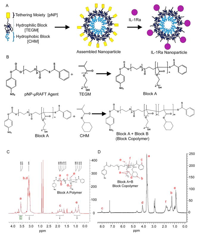
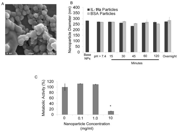
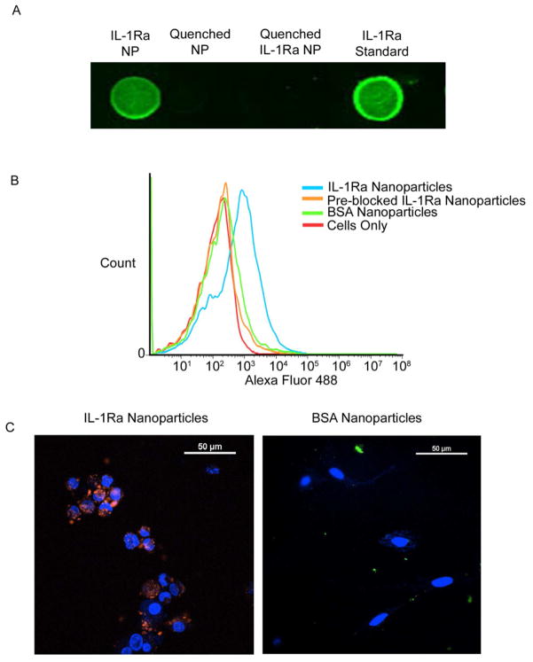
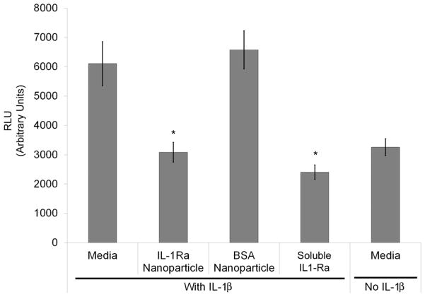
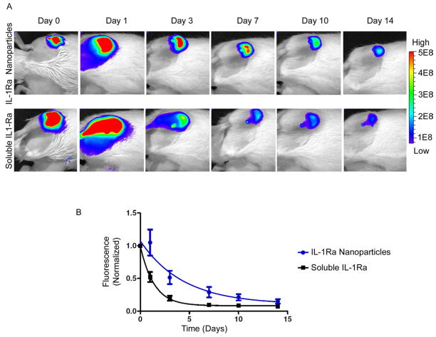
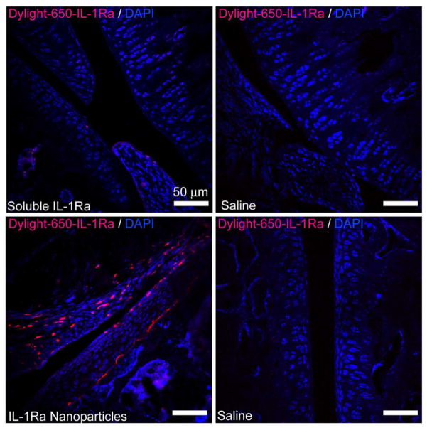
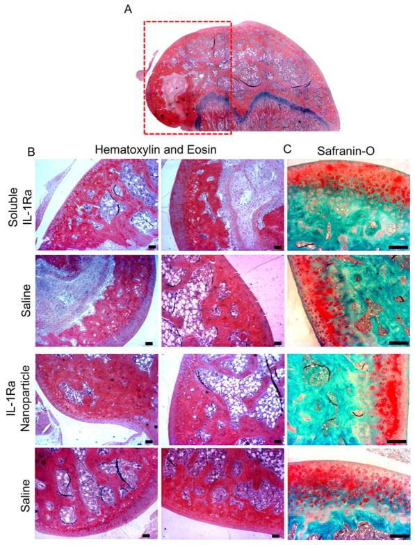
Intra-articular delivery of therapeutics to modulate osteoarthritis (OA) is challenging. Delivery of interleukin-1 receptor antagonist (IL-1Ra), the natural protein inhibitor of IL-1, to modulate IL-1-based inflammation through gene therapy or bolus protein injections has emerged as a promising therapy for OA. However, these approaches suffer from rapid clearance and reduced potency over time. Nano/microparticles represent a promising strategy for overcoming the shortcomings of intra-articular drug delivery. However, these delivery vehicles are limited for delivery of protein therapeutics due to their hydrophobic character, low drug loading efficiency, and harsh chemical conditions during particle processing. We designed a new block copolymer that assembles into submicron-scale particles and provides for covalently tethering proteins to the particle surface for controlled intra-articular protein delivery. This block copolymer self-assembles into 300 nm-diameter particles with a protein tethering moiety for surface covalent conjugation of IL-1Ra protein. This copolymer particle system efficiently bound IL-1Ra and maintained protein bioactivity in vitro. Furthermore, particle-tethered IL-1Ra bound specifically to target synoviocyte cells via surface IL-1 receptors. Importantly, IL-1Ra nanoparticles inhibited IL-1-mediated signaling to equivalent levels as soluble IL-1Ra. Finally, the ability of nanoparticles to retain IL-1Ra in the rat stifle joint was evaluated by in vivo imaging over 14 days. IL-1Ra-tethered nanoparticles significantly increased the retention time of IL-1Ra in the rat stifle joint over 14 days with enhanced IL-1Ra half-life (3.01 days) compared to that of soluble IL-1Ra (0.96 days) and without inducing degenerative changes in cartilage structure or composition.


Figures

Figure 1
Figure 1. Protein-tethering block copolymer
A) Schematic of self-assembly of the nanoparticles and protein conjugation. B) A modified commercial RAFT agent (μ-RAFT) was used for polymerization. Tetraethylene glycol methacrylate and μ-RAFT were mixed and polymerization was initiated using AIBN. Monomeric cyclohexyl methacrylate was added to the product of the first reaction (block A) and polymerization was re-initiated to form the copolymer (block A+B). C) The synthesis of the copolymer is confirmed by 1H-NMR. The block A polymer synthesis was confirmed by 1H-NMR. Peaks were observed at 3.7 ppm (14H, 13.98) (c, f), 4.2 ppm (2H, 2.0) (b, e), 1.9 ppm (2H, 1.49) (g), and 0.9–1.1 ppm (3H, 2.56) (a, h). D) The synthesis of the block A+B (copolymer) was confirmed by 1H-NMR. Peaks were observed at 3.6 ppm (m*14H) (a), 4.1 ppm (m*2H) (d), 1.3 ppm (n*10H) (b), 0.9 ppm (m*3H, n*3H) (e), 1.9 ppm (6H) (f), and 7.0 ppm (9H) (c). m = number of TEGM monomers; n = number of CHM monomers.
Figure 2
Figure 2. Block copolymer forms nanoparticles
A) SEM images of nanoparticles. Nanoparticle size was confirmed by scanning electron microscopy. B) Dynamic Light Scattering based sizing of particles. Nanoparticle diameters were measured using a refractive index of 1.33 (water). Nanoparticles were measured after rotary evaporation of solvent (rotovap) referred to as base nanoparticles; after raising the pH to initiate protein tethering (pH > 7.4), and at 15, 30, 45, 60, 120 min, and overnight after adding protein to the particles. C) MTT assay for cytotoxicity for cells incubated with nanoparticles.
Figure 3
Figure 3. Protein tethering and target specificity for IL-1Ra nanoparticles
A) Dot Blot: IL-1Ra-tethered particles, glycine-tethered particles, or soluble IL-1Ra were dried on nitrocellulose membranes and were probed with anti-IL-1Ra antibodies. Blots were imaged using near infrared (800 nm) secondary antibodies and an infrared imager. B) IL-1Ra-tethered nanoparticles bind to synoviocytes in an IL-1R-dependent manner. A synoviocyte cell line (HIG-82) was incubated with fluorescently tagged IL-1Ra-tethered particles or fluorescently tagged BSA-tethered particles, with or without an IL-1β pre-blocking step and assayed using flow cytometry. Synoviocytes + IL-1Ra-Particles (44.0% cells with bound nanoparticles), Blue; Synoviocytes + BSA-Particles (2.7%), Green; Synoviocytes + Pre-Block + IL-1Ra-Particles, (2.8%), Orange; Synoviocytes Only (0.1%), Red. C) Confocal microscopy images showing that IL-1Ra-tethered particles are bound by synoviocytes: Confocal Microscopy Analysis. Particles were incubated with a synoviocyte cell line (HIG-82) for 2 hr. Samples were rinsed three times with PBS before imaging. IL-1Ra-tethered particles, Red; BSA-tethered particles, Green; Nuclei, Blue. 10 fields from 4 samples of each group were analyzed.
Figure 4
Figure 4. IL-1Ra-tethered particles inhibit IL-1β-induced NF-κB activation
NIH 3T3 fibroblasts with an NF-κB-responsive luciferase reporter construct were pre-incubated for 1 h with 1 μg/mL IL-1Ra-tethered particles, BSA-tethered particles, or soluble IL-1Ra before stimulating with 0.1 ng/mL IL-1β for 6 hr. Both IL-1Ra particles and soluble IL-1Ra inhibited NF-κB activation to comparable levels with unstimulated controls (n=3), * p < 0.004.
Figure 5
Figure 5. IL-1Ra-tethered particles are retained longer than soluble IL-1Ra in the intra-articular joint space
A) Representative images of labeled IL-1Ra. IL-1Ra was tagged with a near-IR dye (AF750-maleimide) prior to tethering IL-1Ra to particles. IL-1Ra-tethered particles or soluble IL-1Ra was injected into the right stifle joint of 8–10 wk old rats. Left stifle joints were injected with saline at the same time. Total IR photon counts (relative fluorescence units) within a fixed area centered over the rat’s joint were measured by IVIS imaging over 14 days. B) IL1-Ra-particles show sustained signal compared to soluble IL1-Ra. Infrared (IR) photon counts were measured in each rat over 14 days by an IVIS imaging system. All data were normalized by individual rat to its day 0 photon count. The photon’s signal decay was fit using a one-phase exponential decay model. Half-life: 3.01 ± 0.09 days for IL-1Ra-particles (n = 6) vs. 0.96 ± 0.08 days for soluble IL-1Ra (n = 5).
Figure 6
Figure 6. IL-1Ra-tethered particles are distributed throughout the intra-articular joint space
IL-1Ra was tagged with a Dylight-IR-650 dye prior to tethering IL-1Ra to particles. Tagged IL-1Ra-tethered particles or soluble IL-1Ra was injected into the right stifle joint of 8–10 wk old rats while the left stifle joints received saline. Cryosectioned samples were counterstained with DAPI to localize dye tagged protein. Scale bar = 50 μm.
Figure 7
Figure 7. EPIC-μCT reconstructions of the tibial plateau region cartilage in rats
Representative reconstructions of cartilage from EPIC-μCT analysis. Visual comparisons confirm that there were no gross differences between cartilage receiving IL-1Ra nanoparticles versus soluble IL-1Ra or saline-treated. Rat joints treated with MIA indicated severe cartilage degeneration. Medial side, Left; Lateral side, Right.
Figure 8
Figure 8. Histological sections indicating no alterations in cartilage structure and morphology in rats treated with IL-1Ra nanoparticles vs. soluble IL-1Ra or PBS (contralateral)
A) Longitudinal section of distal femur indicating the region of interest (red box). All samples were either stained with B) hematoxylin and eosin staining, or C) sGAG-staining Safranin-O (scale bar = 100 μm).

Similar articles

Cited by

References

    1. CDC. Osteoarthritis. 2011 Available from: http://www.cdc.gov/arthritis/basics/osteoarthritis.htm.
    1. Helmick CG, Felson DT, Lawrence RC, Gabriel S, Hirsch R, Kwoh CK, et al. Estimates of the prevalence of arthritis and other rheumatic conditions in the United States. Part I. Arthritis Rheum. 2008;58:15–25. - PubMed
    1. Peyron JG. Epidemiologic and etiologic approach of osteoarthritis. Semin Arthritis Rheum. 1979;8:288–306. - PubMed
    1. Hellio Le Graverand-Gastineau MP. OA clinical trials: current targets and trials for OA. Choosing molecular targets: what have we learned and where we are headed? Osteoarthritis Cartilage. 2009;17:1393–401. - PubMed
    1. Pelletier JP, Martel-Pelletier J, Abramson SB. Osteoarthritis, an inflammatory disease: potential implication for the selection of new therapeutic targets. Arthritis Rheum. 2001;44:1237–47. - PubMed

Publication types

MeSH terms