Review
. 2012 Aug;18(8):472-82.
doi: 10.1016/j.molmed.2012.06.006. Epub 2012 Jul 18.

Pre-mRNA splicing in disease and therapeutics

Affiliations
Review

Pre-mRNA splicing in disease and therapeutics

Ravi K Singh et al. Trends Mol Med. 2012 Aug.

Abstract

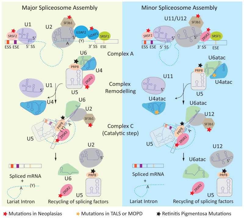
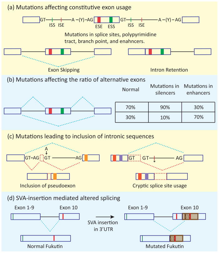
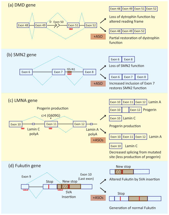
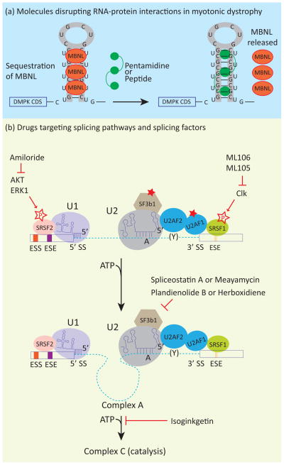
In metazoans, alternative splicing of genes is essential for regulating gene expression and contributing to functional complexity. Computational predictions, comparative genomics, and transcriptome profiling of normal and diseased tissues indicate that an unexpectedly high fraction of diseases are caused by mutations that alter splicing. Mutations in cis elements cause missplicing of genes that alter gene function and contribute to disease pathology. Mutations of core spliceosomal factors are associated with hematolymphoid neoplasias, retinitis pigmentosa, and microcephalic osteodysplastic primordial dwarfism type 1 (MOPD1). Mutations in the trans regulatory factors that control alternative splicing are associated with autism spectrum disorder, amyotrophic lateral sclerosis (ALS), and various cancers. In addition to discussing the disorders caused by these mutations, this review summarizes therapeutic approaches that have emerged to correct splicing of individual genes or target the splicing machinery.


Figures

Figure 1
Figure 1
Spliceosome assembly and disease-associated mutations in spliceosome components. The dashed lines and rectangles represent introns and exons, respectively. The left panel shows the assembly of the major (U2 type) spliceosome. U1 and U2 snRNPs are recruited to the consensus 5′ splice site (5′ SS) and branch point (A), respectively. The U2-auxiliary factor heterodimer (U2AF2/U2AF1) interacts with the polypyrimidine track (Y) and 3′ splice site (3′ SS), forming complex A. The U4/6 and U5 snRNPs join the assembling spliceosome followed by remodeling of the complex leading to removal of the U1 and U4 snRNP and formation of the catalytic complex (complex C). Two trans-esterification reactions join the exons and release an intron lariat that is subsequently degraded and the spliceosome components are recycled for subsequent rounds of splicing. The right panel shows the assembly of the minor (U12 type) spliceosome, in which U1, U2, U4, and U6 are replaced by homologous U11, U12, U4atac, and U6atac snRNPs, respectively. The red star indicates the components that are mutated in neoplasias. The black star indicates the components that are mutated in retinitis pigmentosa. The orange star indicates the mutation in U4atac that is associated with MOPD1. Abbreviations: ESE, exonic splicing enhancers; ESS, exonic splicing silencers.
Figure 2
Figure 2
Four mechanistic categories of altered gene function by splicing mutations. (a) The basic cis elements of the splicing code are indicated: 5′ and 3′ splice sites (represented by GT and AG), polypyrimidine tract (Y), branch point (A), and exonic and intronic enhancers (ESE and ISE) and silencers (ESS and ISS). Mutations affecting splice sites, the polypyrimidine tract, the branch point, or splicing enhancers lead to exon skipping or intron retention. (b) Mutations in enhancer or silencer elements can change the ratio of isoforms containing alternative exons. (c) Mutations within introns can lead to inclusion of intronic sequences (indicated by red dashed rectangles) by creating a splice site/pseudoexon (indicated by arrow) and/or by creating an enhancer element (indicated by asterisk), allowing recognition of a cryptic splice site. The blue dashed lines indicate the normal splicing pattern, whereas the red dashed lines indicate the splicing pattern caused by the mutation. (d) Insertion of transposable elements (SVAs, represented by a brown rectangle) in the 3′ UTR of the fukutin gene leads to the alternate use of splice sites producing a protein with a different carboxy-terminal sequence (patterned brown rectangle). The green lines indicate the start codon of both the normal and mutated fukutin whereas red lines indicate stop codons.
Figure 3
Figure 3
Use of antisense oligonucleotides for splicing correction therapy. The splicing patterns of the mutated gene in the absence and presence of ASOs are indicated in the top and bottom panels, respectively. The red lines with projections indicate the ASOs. (a) In DMD a deletion of exon 50 (indicated by Δ exon 50) leads to disruption of the reading frame resulting in loss of dystrophin function. ASO-induced skipping of exon 51 restores the reading frame and partially rescues dystrophin function. (b) In SMA, loss of SMN protein from the mutated SMN1 gene can be rescued by inducing inclusion of exon 7 of the SMN2 gene. The use of an ASO targeting an intronic splicing silencers leads to enhanced inclusion of SMN2 exon 7. (c) In Hutchinson-Gilford progeria syndrome, a silent mutation in exon 11, indicated by asterisk labeled c>t (G609G), leads to activation of a cryptic 5′ splice site resulting in the production of a toxic protein, progerin. Combined administration of two ASOs to block the 3′ splice site of exon 10 and the cryptic splice site inhibit progerin production. (d) In FCMD, insertion of an SVA leads to activation of a cryptic 5′ splice site in exon 9 and use of a 3′ splice site in the SVA segment. Combined administration of three ASOs to block the aberrant 3′ splice site and a nearby ESE (shown together), and an ISE near the aberrant 5′ splice site promote use of correct 5′ and 3′ splice sites producing fukutin protein.
Figure 4
Figure 4
(a) Small molecules disrupt aberrant RNA-protein interactions in myotonic dystrophy. Small molecules and peptides have been used to disrupt the interaction between CUG repeat RNA and MBNL. This results in rescue of MBNL splicing functions and reversal of some features of myotonic dystrophy. (b) Several compounds inhibit splicing at various steps of splicing assembly and have potential as antitumorigenic drugs. Meayamycin, a compound derived from spliceostatin A, as well as Pladienolid B and Herboxidiene, bind to the SF3b complex of the U2 snRNP and inhibit the formation of complex A. Isoginkgetin prevents the formation of complex B. Amiloride inhibits Akt1 and Erk1 kinases which phosphorylate SR proteins that are important for regulation of constitutive and alternative splicing. Similarly, ML105 and ML106 were identified in a screen for correction of splicing of the LMNA minigene. These compounds were found to inhibit cdc2-like kinases (Clk) that also phosphorylate SR proteins.

References

    1. Wahl MC, et al. The spliceosome: design principles of a dynamic RNP machine. Cell. 2009;136:701–718. - PubMed
    1. Berget SM. Exon recognition in vertebrate splicing. J Biol Chem. 1995;270:2411–2414. - PubMed
    1. Patel AA, Steitz JA. Splicing double: insights from the second spliceosome. Nat Rev Mol Cell Biol. 2003;4:960–970. - PubMed
    1. Will CL, Luhrmann R. Splicing of a rare class of introns by the U12-dependent spliceosome. Biol Chem. 2005;386:713–724. - PubMed
    1. Nilsen TW, Graveley BR. Expansion of the eukaryotic proteome by alternative splicing. Nature. 2010;463:457–463. - PMC - PubMed