

Heparin-binding epidermal growth factor-like growth factor and mesenchymal stem cells act synergistically to prevent experimental necrotizing enterocolitis
- PMID: 22819639
- PMCID: PMC3444529
- DOI: 10.1016/j.jamcollsurg.2012.05.037
Heparin-binding epidermal growth factor-like growth factor and mesenchymal stem cells act synergistically to prevent experimental necrotizing enterocolitis
Abstract
Background: We have shown that administration of heparin-binding EGF (epidermal growth factor)-like growth factor (HB-EGF) protects the intestines from experimental necrotizing enterocolitis (NEC). We have also demonstrated that systemically administered mesenchymal stem cells (MSC) can engraft into injured intestines. This study investigated the effects of HB-EGF on MSC in vitro, and whether MSC and HB-EGF can act synergistically to prevent NEC in vivo.
Study design: In vitro, the effect of HB-EGF on MSC proliferation, migration, and apoptosis was determined. In vivo, rat pups received MSC either intraperitoneally (IP) or intravenously (IV). Pups were assigned to 1 of 7 groups: Group 1, breast-fed; Group 2, experimental NEC; Group 3, NEC+HB-EGF; Group 4, NEC+MSC IP; Group 5, NEC+HB-EGF+MSC IP; Group 6, NEC+MSC IV; or Group 7, NEC+HB-EGF+MSC IV. Mesechymal stem cell engraftment, histologic injury, intestinal permeability, and mortality were determined.
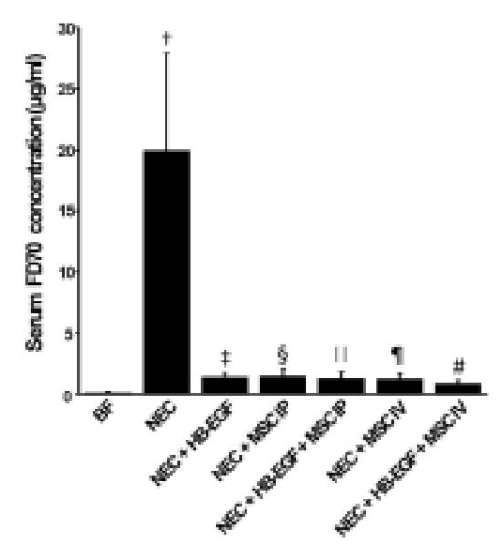
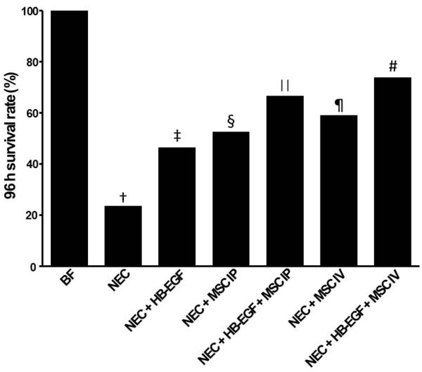
Results: Heparin-binding EGF-like growth factor promoted MSC proliferation and migration, and decreased MSC apoptosis in vitro. In vivo, MSC administered IV had increased engraftment into NEC-injured intestine compared with MSC administered IP (p < 0.05). Heparin binding EGF-like growth factor increased engraftment of IP-administered MSC (p < 0.01) and IV-administered MSC (p < 0.05). Pups in Groups 3 to 7 had a decreased incidence of NEC compared with nontreated pups (Group 2), with the lowest incidence in pups treated with HB-EGF+MSC IV (p < 0.01). Pups in Group 7 had a significantly decreased incidence of intestinal dilation and perforation, and had the lowest intestinal permeability, compared with other treatment groups (p < 0.01). Pups in all experimental groups had significantly improved survival compared with pups exposed to NEC, with the best survival in Group 7 (p < 0.05).
Conclusions: Heparin-binding EGF-like growth factor and MSC act synergistically to reduce injury and improve survival in experimental NEC.
Copyright © 2012 American College of Surgeons. Published by Elsevier Inc. All rights reserved.
Figures


Similar articles
-
Heparin-binding EGF-like growth factor and enteric neural stem cell transplantation in the prevention of experimental necrotizing enterocolitis in mice.Pediatr Res. 2015 Jul;78(1):29-37. doi: 10.1038/pr.2015.63. Epub 2015 Mar 25. Pediatr Res. 2015. PMID: 25806717 Free PMC article.
-
Heparin-binding epidermal growth factor-like growth factor promotes enterocyte migration and proliferation in neonatal rats with necrotizing enterocolitis.J Pediatr Surg. 2007 Jan;42(1):214-20. doi: 10.1016/j.jpedsurg.2006.09.055. J Pediatr Surg. 2007. PMID: 17208569
-
Heparin-binding EGF-like growth factor increases intestinal microvascular blood flow in necrotizing enterocolitis.Gastroenterology. 2009 Jul;137(1):221-30. doi: 10.1053/j.gastro.2009.03.060. Epub 2009 Apr 8. Gastroenterology. 2009. PMID: 19361505 Free PMC article.
-
Heparin-binding EGF-like growth factor (HB-EGF) and necrotizing enterocolitis.Semin Pediatr Surg. 2005 Aug;14(3):167-74. doi: 10.1053/j.sempedsurg.2005.05.005. Semin Pediatr Surg. 2005. PMID: 16084404 Review.
-
Milk epidermal growth factor and gut protection.J Pediatr. 2010 Feb;156(2 Suppl):S31-5. doi: 10.1016/j.jpeds.2009.11.018. J Pediatr. 2010. PMID: 20105663 Free PMC article. Review.
Cited by
-
Stem cell therapy as a promising strategy in necrotizing enterocolitis.Mol Med. 2022 Sep 6;28(1):107. doi: 10.1186/s10020-022-00536-y. Mol Med. 2022. PMID: 36068527 Free PMC article. Review.
-
Treatment of experimental necrotizing enterocolitis with stem cell-derived exosomes.J Pediatr Surg. 2018 Jun;53(6):1215-1220. doi: 10.1016/j.jpedsurg.2018.02.086. Epub 2018 Mar 14. J Pediatr Surg. 2018. PMID: 29661576 Free PMC article.
-
New insights into necrotizing enterocolitis: From laboratory observation to personalized prevention and treatment.J Pediatr Surg. 2019 Mar;54(3):398-404. doi: 10.1016/j.jpedsurg.2018.06.012. Epub 2018 Jun 18. J Pediatr Surg. 2019. PMID: 29980346 Free PMC article. Review.
-
Enteric nervous system abnormalities are present in human necrotizing enterocolitis: potential neurotransplantation therapy.Stem Cell Res Ther. 2013;4(6):157. doi: 10.1186/scrt387. Stem Cell Res Ther. 2013. PMID: 24423414 Free PMC article.
-
Cell-based therapies in preclinical models of necrotizing enterocolitis: a systematic review and meta-analysis.Stem Cells Transl Med. 2025 Feb 11;14(2):szae102. doi: 10.1093/stcltm/szae102. Stem Cells Transl Med. 2025. PMID: 40036304 Free PMC article.
References
-
- Kliegman RM, Fanaroff AA. Necrotizing enterocolitis. N Engl J Med. 1984;310:1093–1103. - PubMed
-
- Feng J, El-Assal ON, Besner GE. Heparin-binding epidermal growth factor-like growth factor decreases the incidence of necrotizing enterocolitis in neonatal rats. J Pediatr Surg. 2006;41:144–149. discussion 144-149. - PubMed
-
- Feng J, El-Assal ON, Besner GE. Heparin-binding EGF-like growth factor (HB-EGF) and necrotizing enterocolitis. Semin Pediatr Surg. 2005;14:167–174. - PubMed
MeSH terms
Substances
Grants and funding
LinkOut - more resources
Full Text Sources
