

Tissue plasminogen activator treatment of stroke in type-1 diabetes rats
- PMID: 22820263
- PMCID: PMC3474540
- DOI: 10.1016/j.neuroscience.2012.07.018
Tissue plasminogen activator treatment of stroke in type-1 diabetes rats
Abstract
Background and purpose: Diabetes mellitus (DM) is a major stroke risk factor and is associated with poor recovery compared with nondiabetic stroke patients. In the present study, we investigated the effects of tissue plasminogen activator (tPA) treatment of stroke in diabetic and non-diabetic rats.
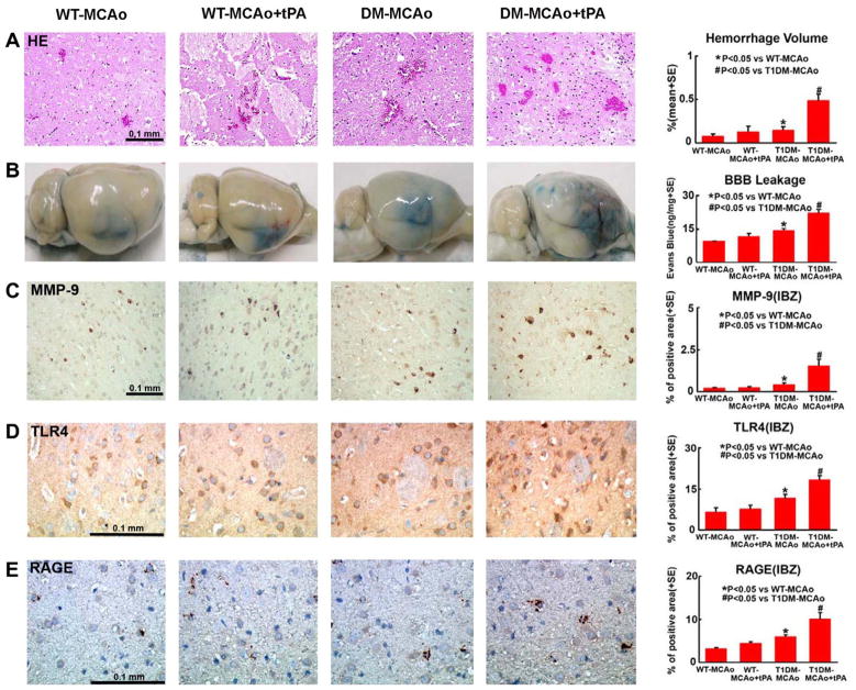
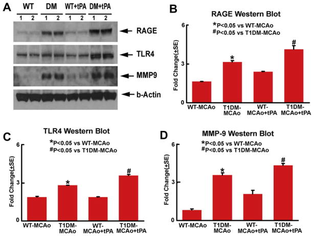
Methods: Type-1 diabetes (T1DM) was induced by injection of streptozotocin. Non-T1DM and T1DM rats were subjected to embolic middle cerebral artery occlusion (MCAo) and treated with or without tPA 2h after MCAo. Functional outcomes and immunostaining for advanced glycation endproducts receptor (RAGE), matrix metalloproteinase-9 (MMP-9) and toll-like receptor 4 (TLR4) and Western blotting were performed.
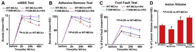
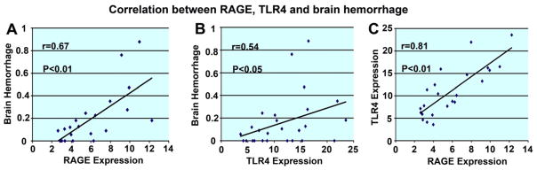
Results: tPA treatment of WT-MCAo rats significantly improved the functional outcome and reduced the lesion volume compared with non-treatment WT-MCAo rats (p<0.05). There was no significant difference between treatment with or without tPA in the WT-MCAo group in brain hemorrhage, BBB leakage and expression of inflammatory mediators, RAGE, MMP-9 and TLR4. However, tPA treatment in T1DM-MCAo rats (T1DM-MCAo+tPA) significantly enlarged brain hemorrhage, augmented BBB leakage, and failed to decrease lesion volume and improve functional outcome after stroke compared to T1DM-MCAo control. tPA treatment also significantly increased the expression of RAGE, MMP-9 and TLR4 in the ischemic brain in T1DM-MCAo rats compared with T1DM-MCAo control rats (p<0.05). Brain hemorrhage was significantly correlated with functional deficit and RAGE and TLR4 expression, respectively.
Conclusions: Treatment of stroke with tPA increased brain hemorrhage, BBB leakage and failed to improve functional outcome in T1DM rats. The increased inflammatory response may contribute to the failed neuroprotective effects of tPA treatment in T1DM rats.
Copyright © 2012 IBRO. Published by Elsevier Ltd. All rights reserved.
Figures
Similar articles
-
Neamine induces neuroprotection after acute ischemic stroke in type one diabetic rats.Neuroscience. 2014 Jan 17;257:76-85. doi: 10.1016/j.neuroscience.2013.10.071. Epub 2013 Nov 8. Neuroscience. 2014. PMID: 24211797 Free PMC article.
-
Niaspan reduces high-mobility group box 1/receptor for advanced glycation endproducts after stroke in type-1 diabetic rats.Neuroscience. 2011 Sep 8;190:339-45. doi: 10.1016/j.neuroscience.2011.06.004. Epub 2011 Jun 13. Neuroscience. 2011. PMID: 21683770 Free PMC article.
-
Angiopoietin-1 Mimetic Peptide Promotes Neuroprotection after Stroke in Type 1 Diabetic Rats.Cell Transplant. 2018 Dec;27(12):1744-1752. doi: 10.1177/0963689718791568. Epub 2018 Aug 20. Cell Transplant. 2018. PMID: 30124060 Free PMC article.
-
Tissue plasminogen activator (tPA) and matrix metalloproteinases in the pathogenesis of stroke: therapeutic strategies.CNS Neurol Disord Drug Targets. 2008 Jun;7(3):243-53. doi: 10.2174/187152708784936608. CNS Neurol Disord Drug Targets. 2008. PMID: 18673209 Free PMC article. Review.
-
Efficacy of Chinese herbal medicine for tPA thrombolysis in experimental stroke: A systematic review and meta-analysis.Phytomedicine. 2022 Jun;100:154072. doi: 10.1016/j.phymed.2022.154072. Epub 2022 Mar 23. Phytomedicine. 2022. PMID: 35349833
Cited by
-
Ischemic stroke and diabetes: a TLR4-mediated neuroinflammatory perspective.J Mol Med (Berl). 2024 Jun;102(6):709-717. doi: 10.1007/s00109-024-02441-9. Epub 2024 Mar 28. J Mol Med (Berl). 2024. PMID: 38538987 Review.
-
Acute Hyperglycemia Exacerbates Hemorrhagic Transformation after Embolic Stroke and Reperfusion with tPA: A Possible Role of TXNIP-NLRP3 Inflammasome.J Stroke Cerebrovasc Dis. 2022 Feb;31(2):106226. doi: 10.1016/j.jstrokecerebrovasdis.2021.106226. Epub 2021 Nov 27. J Stroke Cerebrovasc Dis. 2022. PMID: 34847489 Free PMC article.
-
Exacerbated VEGF up-regulation accompanies diabetes-aggravated hemorrhage in mice after experimental cerebral ischemia and delayed reperfusion.Neural Regen Res. 2022 Jul;17(7):1566-1575. doi: 10.4103/1673-5374.330612. Neural Regen Res. 2022. PMID: 34916442 Free PMC article.
-
Inhibition of Toll-Like Receptor-4 (TLR-4) Improves Neurobehavioral Outcomes After Acute Ischemic Stroke in Diabetic Rats: Possible Role of Vascular Endothelial TLR-4.Mol Neurobiol. 2019 Mar;56(3):1607-1617. doi: 10.1007/s12035-018-1184-8. Epub 2018 Jun 16. Mol Neurobiol. 2019. PMID: 29909454 Free PMC article.
-
Diabetes mellitus aggravates hemorrhagic transformation after ischemic stroke via mitochondrial defects leading to endothelial apoptosis.PLoS One. 2014 Aug 18;9(8):e103818. doi: 10.1371/journal.pone.0103818. eCollection 2014. PLoS One. 2014. PMID: 25133692 Free PMC article.
References
-
- Albers GW, Bates VE, Clark WM, Bell R, Verro P, Hamilton SA. Intravenous tissue-type plasminogen activator for treatment of acute stroke: the Standard Treatment with Alteplase to Reverse Stroke (STARS) study. JAMA. 2000;283:1145–1150. - PubMed
-
- Alvarez-Sabin J, Molina CA, Montaner J, Arenillas JF, Huertas R, Ribo M, Codina A, Quintana M. Effects of admission hyperglycemia on stroke outcome in reperfused tissue plasminogen activator-treated patients. Stroke. 2003;34:1235–1241. - PubMed
-
- Barile GR, Schmidt AM. RAGE and its ligands in retinal disease. Curr Mol Med. 2007;7:758–765. - PubMed
-
- Barone FC, Feuerstein GZ. Inflammatory mediators and stroke: new opportunities for novel therapeutics. J Cereb Blood Flow Metab. 1999;19:819–834. - PubMed
-
- Barth TM, Grant ML, Schallert T. Effects of MK-801 on recovery from sensorimotor cortex lesions. Stroke. 1990;21:III153–III157. - PubMed
Publication types
MeSH terms
Substances
Grants and funding
LinkOut - more resources
Full Text Sources
Medical
Miscellaneous
