. 2012 Aug;138(2):179-86.
doi: 10.1007/s00418-012-0999-z. Epub 2012 Jul 22.

Formation and migration of neural crest cells in the vertebrate embryo

Affiliations

Formation and migration of neural crest cells in the vertebrate embryo

Marianne E Bronner. Histochem Cell Biol. 2012 Aug.

Abstract

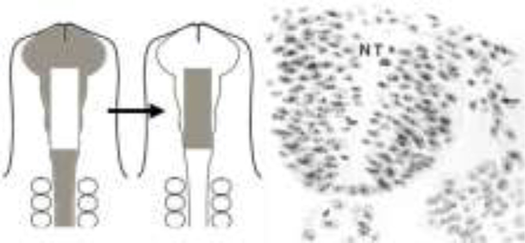
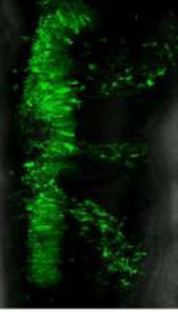
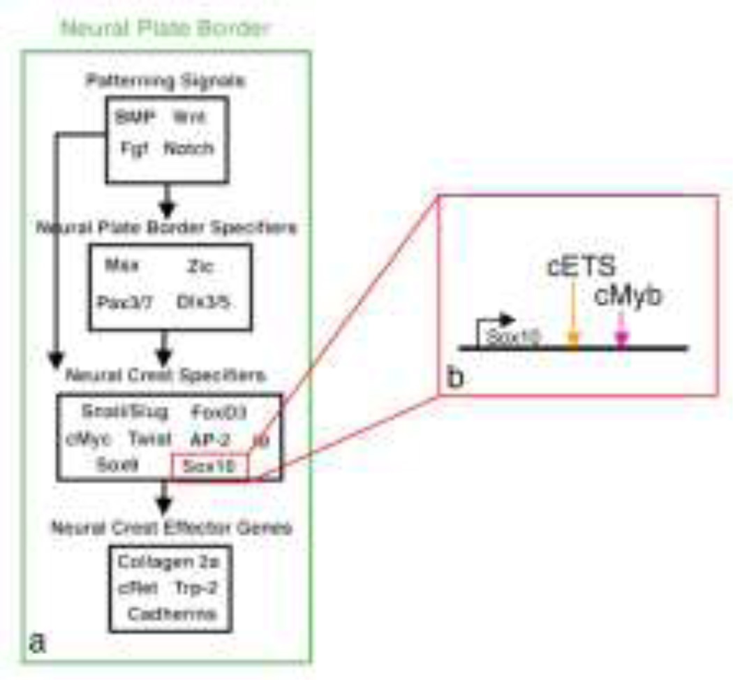
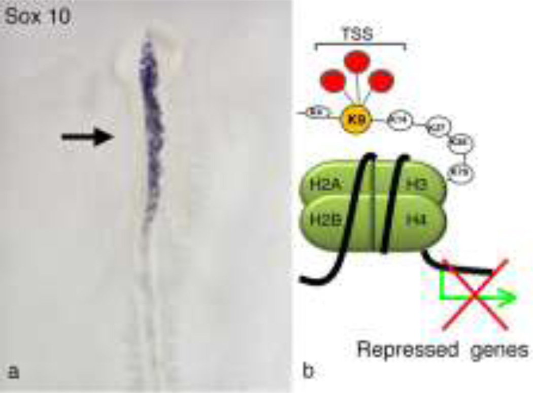
The neural crest is a stem cell population, unique to vertebrates, that gives rise to a vast array of derivatives, ranging from peripheral ganglia to the facial skeleton. This population is induced in the early embryo at the border of the neural plate, which will form the central nervous system (CNS). After neural tube closure, neural crest cells depart from the dorsal CNS via an epithelial to mesenchymal transition (EMT), forming a migratory mesenchymal cell type that migrates extensive to diverse locations in the embryo. Using in vivo loss-of-function approaches and cis-regulatory analysis coupled with live imaging, we have investigated the gene regulatory network that mediates formation of this fascinating cell type. The results show that a combination of transcriptional inputs and epigenetic modifiers control the timing of onset of neural crest gene expression. This in turn leads to the EMT process that produces this migratory cell population.


Figures

Fig. 1
Fig. 1
Schematic diagram illustrating the process of neural crest formation. At gastrulation stages (top panel), the neuroectoderm is a flat neural plate (future central nervous system) that apposes the non neural ectoderm (future epidermis). The neural plate border (purple) contains presumptive neural crest cells. As neurulation proceeds (middle panel), the neural folds (purple) have risen, and from the neural plate which will close to form the neural tube. After neural tube closure (bottom panel), neural crest cells undergo an epithelial to mesenchymal transition and migrate from the neural tube into the peripheral as individual mesenchymal cells. At trunk levels illustrated here, they migrate around the somites with those cells migrating dorsally above the somites forming melanocytes whereas those migrating ventrally forming sensory and sympathetic ganglia.
Fig. 2
Fig. 2
Quail-chick chimera. Diagram showing how neural folds from quail (gray) are grafted into a chick host. Section through at embryo showing Feulgen stained quail cells (arrow) migrating into the periphery after grafting into chick host. (adapted from Bronner and LeDouarin, 2012).
Fig. 3
Fig. 3
A frame of a time lapse moving from an embryo imaged after a construct encoding green fluorescent protein (GFP) was electroporated into the right side of the neural tube. Because neural crest cells are the only cells to migrate from the neural tube, it is possible to follow their migration in real time from the neural tube to the periphery.
Fig. 4
Fig. 4
(a) A putative gene regulatory network helps to explain the molecular progression of neural crest formation. First, inductive patterning signals, including BMPs, Wnts, and FGFs cooperate to induce the neural plate border containing cells with neural crest potential. These signals in turn up-regulate a suite of genes at the neural plate border, including Pax, Zic, and Msx genes. The neural plate border genes in turn regulate neural crest specifier genes, like Sox10, FoxD3 and Snail2. These help to mediate neural crest EMT by regulating cadherins as well as numerous differentiation genes. (b) Cis-regulatory analysis of Sox10 and other genes are helping to expand the NC-GRN by identifying new inputs (e.g. Ets1 and Myb transcription factors) as well as demonstrating direct regulatory interactions. The newly identified neural crest enhancers also serve as useful tools for imaging, and cell type specific over-expression and knock-down.
Fig. 5
Fig. 5
(a) Morpholino knock-down of the histone demethylase JmjD2A causes loss of the neural crest specifier gene Sox10 on the electroporated (black arrow) side, whereas there is normal expression on the control, non-electroporated side. (b) Schematic diagram shows the repressive state when histone H3 is trimethylated on lysine 9. JmjD2A specifically removes this repressive mark, thereby allowing onset of Sox10 transcription at the appropriate stages. After morpholino-mediated loss of JmjD2A, however, the repressive mark remains on and Sox10 transcription is inhibited. (Data from Strobl-Mazzulla et al., 2010).

References

    1. Adams M, Gammill L, Bronner-Fraser M. Discovery of transcription factors and other candidate regulators of neural crest development. Dev Dynamics. 2008;237:1021–1033. - PMC - PubMed
    1. Basch ML, Bronner-Fraser M, Garcia-Castro MI. Specification of the neural crest occurs during gastrulation and requires Pax7. Nature. 2006;441:218–222. - PubMed
    1. Betancur P, Bronner-Fraser M, Sauka-Spengler T. Genomic code for Sox10, a key regulatory enhancer for cranial neural crest. Proc Natl Acad Sci USA. 2010a;107:3570–3575. - PMC - PubMed
    1. Betancur P, Bronner-Fraser M, Sauka-Spengler T. Assembling neural crest regulatory circuits into a gene regulatory network. Annu Rev Cell Dev Biol. 2010b;26:581–603. - PMC - PubMed
    1. Bronner ME, LeDoaurin NM. Development and evolution of the neural crest: An overview. Dev Biol. 2010;366:2–9. - PMC - PubMed

Publication types

LinkOut - more resources