Review
. 2012 Oct;29(10):1251-66.
doi: 10.1039/c2np20020a. Epub 2012 Jul 23.

Diversity of P450 enzymes in the biosynthesis of natural products

Affiliations
Review

Diversity of P450 enzymes in the biosynthesis of natural products

Larissa M Podust et al. Nat Prod Rep. 2012 Oct.

Abstract

Diverse oxygenation patterns of natural products generated by secondary metabolic pathways in microorganisms and plants are largely achieved through the tailoring reactions catalysed by cytochrome P450 enzymes (P450s). P450s are a large family of oxidative hemoproteins found in all life forms from prokaryotes to humans. Understanding the reactivity and selectivity of these fascinating C-H bond-activating catalysts will advance their use in generating valuable pharmaceuticals and products for medicine, agriculture and industry. A major strength of this P450 group is its set of established enzyme-substrate relationships, the source of the most detailed knowledge on how P450 enzymes work. Engineering microbial-derived P450 enzymes to accommodate alternative substrates and add new functions continues to be an important near- and long-term practical goal driving the structural characterization of these molecules. Understanding the natural evolution of P450 structure-function should accelerate metabolic engineering and directed evolutionary approaches to enhance diversification of natural product structures and other biosynthetic applications.


Figures

Fig. 1
Fig. 1. Phylogenic diversity of structurally defined bacterial natural product P450s
Tree is based on the sequence alignments in Fig. 5, and built using PhyloWidget, a web-based visualization tool
Fig. 2
Fig. 2. Albaflavenone and farnesene biosynthetic pathway in S. coelicolor A3(2)
Fig. 3
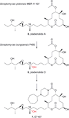
Fig. 3. Pladienolide natural products
Conversion of pladienolide A to semi-synthetic analog E7107 by an engineered strain of S. platensis bearing a P450 gene from S. bungoensis.
Fig. 4
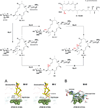
Fig. 4. PikC structure in open and closed conformations
A, P450 protein scaffold is built around a four-helix bundle core (enclosed in red box). Transition between open and closed conformations is approximated linearly by an ensemble of conformers generated computationally and superimposed with the experimentally obtained open and closed PikC structures. Molecule is viewed along the L-helix harbouring the invariant cysteine residue at the N-terminus facing the viewer. Ribbon representation of the open (B) and closed (C) conformations as observed in ligand-free PikC. The mobile F- and G-helices are highlighted in pink, I-helix in green, β-sheets in yellow; heme is shown as green sticks. Molecules in B and C are rotated ~90° toward the viewer along the horizontal axis in the plane of the figure as compared to A.
Fig. 5
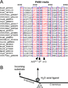
Fig. 5. Sequence alignments
A, Sequence alignments encompassing the I-helix of natural product P450s. Alignment was performed using NCBI’s COBALT-constraint-based multiple alignment online tool. UniProt database (http://www.uniprot.org/) accession numbers are provided for each protein sequence. Residue numbering in (A) corresponds to EryF. B, Cartoon illustrating putative coupling mechanics of the substrate entry and release of the iron axial water ligand.
Fig. 6
Fig. 6. P450 oxygen scission site
Hydrophobicity of the active site in CYP130 increases as a result of the Gn−1 to alanine mutation (A) compared to the wild type enzyme (B). Water molecules bound in the active site are shown in red spheres, heme is in yellow sticks and protein is in blue sticks. Interactions with an axial water ligand are indicated by dashed lines. G243A substitution in the active site also facilitates expulsion of axial water ligand by incoming inhibitors.
Fig. 7
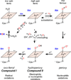
Fig. 7. Conventional P450 catalytic cycle
RH represents the substrate and ROH the resulting monooxygenated product. RO and ROOH depict singe oxygen atom donors, oxotransfer agents and organic peroxides, respectively. Shunt and uncoupling pathways are indicated with dashed-line arrows.
Fig. 8
Fig. 8. Substrate assisted catalysis in EryF (A) and PimD (B)
A, 6-deoxyerythronolide B (yellow sticks) in the catalytic site of EryF. Oxygen molecule bound to the heme iron is shown as a red stick. B, 4,5-desepoxypimaricin (yellow sticks) in the catalytic site of PimD. A fragment of the electron density map (blue mesh) indicates inward rotation of the S238 side chain toward the I-helix groove. Fe axial water ligand is shown as red sphere. H-bonding interactions are indicated by dashed lines with distances in angstroms. Heme is in orange spheres. Fragments of the I-helix are shown as a grey ribbon or green sticks.
Fig. 9
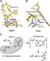
Fig. 9. Distinct epoxidation mechanisms in EpoK and PimD
Epoxidation product epothilone B (yellow) is shown bound in the EpoK active site (A) and a fragment of substrate desepoxypimaricin (yellow) in the PimD active site (B). Blue arrow points are collinear with the π-orbitals in the double bonds C12–C13 in epothilone and C4–C5 in desepoxypimaricin. Fragment of the I-helix is shown as a gray ribbon. Distances are in angstroms. C, PimD epoxidation reaction scheme. Substrate atoms are outlined in grey.
Fig. 10
Fig. 10. Mechanism of aryl-aryl coupling of chromopyrrolic acid by StaP
Fig. 11
Fig. 11. Carrier protein-assisted (A) and chemical group-assisted (B) substrate delivery
A, Complex between P450BioI (cyan) and the acyl carrier protein (green). Fatty acid substrate (yellow sticks) assumes U-shape in the active site. For clarity, the P450 F-, G- and I-helices are highlighted in pink, blue and light green, respectively. B, Multiple binding modes of desosaminyl cycloalkane (pink and cyan) in the active site of PikC. PikC side chains are in green, heme in orange.
Fig. 12
Fig. 12. Oxidative reactions of P450 TamI
Four consecutive oxidation steps catalyzed by P450 TamI and flavoprotein TamL in the biosynthesis of tirandamycin B in Streptomyces sp. 307-9.
Fig. 13
Fig. 13. Substrate binding in MycG
A, Substrates M-IV (34) and M-V (35) (yellow sticks) are bound orthogonal to the heme plane with mycinose methoxy groups at van der Waals distances to the heme (green spheres), preventing access of C-14 and C12–C13 double bond to the Fe centre. Distances between Fe and two reactive centres are in angstroms. B, Javose-decorated precursor, M-III (38), in the parallel orientation presenting the wrong side of the macrolactone ring to the heme. Electron density for M-III is shown in blue mesh.
Fig. 14
Fig. 14. Oxidative reactions of fungal P450 Tir4
Four consecutive oxidation steps catalysed by Tir4 in the biosynthesis of trichothecenes in fungi F. graminearum.

References

    1. Poulos TL, Finzel BC, Gunsalus IC, Wagner GC, Kraut J. J. Biol. Chem. 1985;260:16122–16130. - PubMed
    1. Poulos TL, Finzel BC, Howard AJ. Biochemistry. 1986;25:5314–5322. - PubMed
    1. Poulos TL, Finzel BC, Howard AJ. J. Mol. Biol. 1987;195:687–700. - PubMed
    1. Poulos TL, Howard AJ. Biochemistry. 1987;26:8165–8174. - PubMed
    1. Cupp-Vickery J, Anderson R, Hatziris Z. Proc. Natl. Acad. Sci. U S A. 2000;97:3050–3055. - PMC - PubMed

Publication types

Substances