

Culture, characterization and differentiation of cells from buffalo (Bubalus bubalis) amnion
- PMID: 22820992
- PMCID: PMC3536876
- DOI: 10.1007/s10616-012-9464-z
Culture, characterization and differentiation of cells from buffalo (Bubalus bubalis) amnion
Abstract
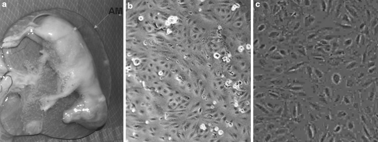
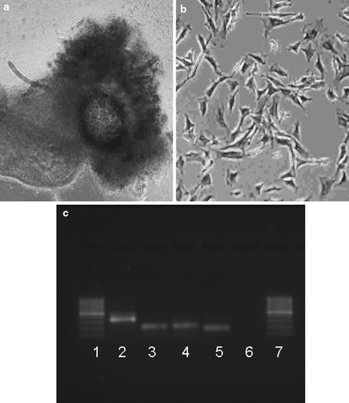
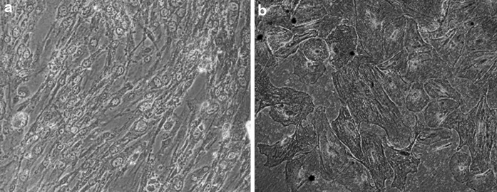
Stem cells present an important tool in livestock assisted reproduction and veterinary therapeutic field such as tissue engineering. We report for the first time isolation of pluripotent stem cell-like cells expressing pluripotency markers (alkaline phospahatase, OCT-4, NANOG and SOX-2) from the amnion of water buffalo (Bubalus bubalis). The cells showed no apparent abnormalities in their chromosomal profiles before and after cryopreservation. The cytochemical staining revealed that pluripotent cells were capable of undergoing directed differentiation in vitro into osteocytes. It could be inferred that amnion-derived pluripotent stem cell-like cells can be isolated, cultured for many passages and differentiated into mesoderm lineage, and may be an alternative source to mesenchymal stem cells. These cells can have applications in assisted reproduction, developmental biological and regenerative medicine.
Figures
Similar articles
-
Buffalo (Bubalus bubalis) term amniotic-membrane-derived cells exhibited mesenchymal stem cells characteristics in vitro.In Vitro Cell Dev Biol Anim. 2015 Oct;51(9):915-21. doi: 10.1007/s11626-015-9920-0. Epub 2015 May 28. In Vitro Cell Dev Biol Anim. 2015. PMID: 26019121
-
Expression of pluripotency genes in buffalo (Bubalus bubalis) amniotic fluid cells.Reprod Domest Anim. 2011 Aug;46(4):705-11. doi: 10.1111/j.1439-0531.2010.01733.x. Epub 2010 Dec 30. Reprod Domest Anim. 2011. PMID: 21198969
-
Isolation and differentiation potential of an equine amnion-derived stromal cell line.Cytotechnology. 2012 Jan;64(1):1-7. doi: 10.1007/s10616-011-9398-x. Epub 2011 Oct 13. Cytotechnology. 2012. PMID: 21994048 Free PMC article.
-
The domesticated buffalo - An emerging model for experimental and therapeutic use of extraembryonic tissues.Theriogenology. 2020 Jul 15;151:95-102. doi: 10.1016/j.theriogenology.2020.04.003. Epub 2020 Apr 7. Theriogenology. 2020. PMID: 32320839 Review.
-
Human amnion-derived cells as a reliable source of stem cells.Curr Mol Med. 2012 Dec;12(10):1340-9. doi: 10.2174/156652412803833625. Curr Mol Med. 2012. PMID: 23016591 Review.
Cited by
-
In-vitro meat: a promising solution for sustainability of meat sector.J Anim Sci Technol. 2021 Jul;63(4):693-724. doi: 10.5187/jast.2021.e85. Epub 2021 Jul 31. J Anim Sci Technol. 2021. PMID: 34447949 Free PMC article. Review.
-
Stem cell therapies and benefaction of somatic cell nuclear transfer cloning in COVID-19 era.Stem Cell Res Ther. 2021 May 12;12(1):283. doi: 10.1186/s13287-021-02334-5. Stem Cell Res Ther. 2021. PMID: 33980321 Free PMC article. Review.
-
Buffalo (Bubalus bubalis) term amniotic-membrane-derived cells exhibited mesenchymal stem cells characteristics in vitro.In Vitro Cell Dev Biol Anim. 2015 Oct;51(9):915-21. doi: 10.1007/s11626-015-9920-0. Epub 2015 May 28. In Vitro Cell Dev Biol Anim. 2015. PMID: 26019121
-
A Comparative Study of Growth Kinetics, In Vitro Differentiation Potential and Molecular Characterization of Fetal Adnexa Derived Caprine Mesenchymal Stem Cells.PLoS One. 2016 Jun 3;11(6):e0156821. doi: 10.1371/journal.pone.0156821. eCollection 2016. PLoS One. 2016. PMID: 27257959 Free PMC article.
-
Isolation and characterization of buffalo (bubalus bubalis) amniotic mesenchymal stem cells derived from amnion from the first trimester pregnancy.J Vet Med Sci. 2018 Apr 27;80(4):710-719. doi: 10.1292/jvms.17-0556. Epub 2018 Mar 8. J Vet Med Sci. 2018. PMID: 29515060 Free PMC article.
References
-
- Anand T, Kumar D, Singh MK, Shah RA, Chauhan MS, Manik RS, Singla SK, Palta P. Buffalo (Bubalus bubalis) embryonic stem cell-like cells and preimplantation embryos exhibit comparable expression of pluripotency-related antigens. Reprod Domest Anim. 2011;46:50–58. doi: 10.1111/j.1439-0531.2009.01564.x. - DOI - PubMed
-
- Bailo M, Soncini M, Vertua E, Signoroni PB, Sanzone S, Lombardi G, Arienti D, Calamani F, Zatti D, Paul P, Albertini A, Zorzi F, Cavagnini A, Candotti F, Wengler GS, Parolini O. Engraftment potential of human amnion and chorion cells derived from term placenta. Transplantation. 2004;78:1439–1448. doi: 10.1097/01.TP.0000144606.84234.49. - DOI - PubMed
LinkOut - more resources
Full Text Sources
Research Materials
Miscellaneous
