Review
. 2012 Sep;43(3):1087-108.
doi: 10.1007/s00726-012-1289-8. Epub 2012 Jul 22.

Current challenges in software solutions for mass spectrometry-based quantitative proteomics

Affiliations
Review

Current challenges in software solutions for mass spectrometry-based quantitative proteomics

Salvatore Cappadona et al. Amino Acids. 2012 Sep.

Abstract

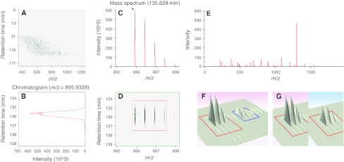
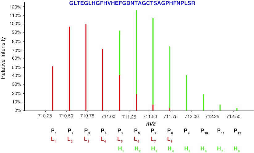
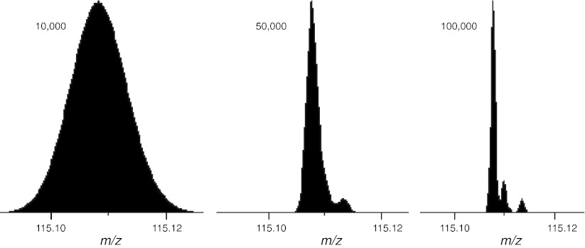
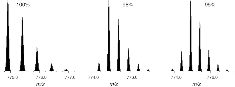
Mass spectrometry-based proteomics has evolved as a high-throughput research field over the past decade. Significant advances in instrumentation, and the ability to produce huge volumes of data, have emphasized the need for adequate data analysis tools, which are nowadays often considered the main bottleneck for proteomics development. This review highlights important issues that directly impact the effectiveness of proteomic quantitation and educates software developers and end-users on available computational solutions to correct for the occurrence of these factors. Potential sources of errors specific for stable isotope-based methods or label-free approaches are explicitly outlined. The overall aim focuses on a generic proteomic workflow.


Figures

Fig. 1
Fig. 1
LC–MS signals. a The ion intensity map gives a bird-eye view of the whole LC–MS experiment. Highlighted in green is a peptide feature magnified in panel d. b Extracted ion chromatogram (XIC) of the monoisotopic peak of the selected peptide ion. The signal shows the ion intensity as a function of the elution time. The area under the curve (AUC) represents the total signal of the monoisotopic peak. c Mass spectrum of the selected peptide ion at maximum chromatographic intensity. The m/z difference of 0.5 Th between contiguous isotopic peaks allows deriving a charge state of 2. The arrow indicates the monoisotopic peak. d Ion intensity map of the peptide ion of interest. The green cross indicates the precursor ion selected for fragmentation. e Tandem mass spectrum of the monoisotopic peak of the selected peptide ion, highlighted by a green cross in panel d. The mass difference between selected peaks allows deriving the amino acids sequence. f For stable isotope-based quantification peptides from two different samples are detected in the same LC–MS run at a characteristic mass difference. g For label-free quantification corresponding peptides from two different samples are detected at the same mass and similar retention time in two different LC–MS runs
Fig. 2
Fig. 2
Overlapping isotopic clusters. Isotopic distribution of the dimethyl labeled peptide GLTEGLHGFHVHEFGDNTAGCTSAGPHFNPLSR. The mass shift of the two isotopologues is smaller than their isotopic envelope, resulting in the overlap of the fifth and consecutive peaks of the light peptide on the monoisotopic and consecutive peaks of the heavy one. In this example, the two peptides are equally abundant, but a quantification strategy that evaluates peptide ratios based on their monoisotopic peaks would largely overestimate the heavy peptide
Fig. 3
Fig. 3
Theoretical iTRAQ data in the region around the 115 reporter ion at different resolutions. The model assumes 95 % purity for 13C and 15N and a 1:1:1:1 mixing ratio. The three peaks seen at a resolution of 100,000 FWHM are (from left to right) the monoisotopic 115 peak, the peak from the partial enrichment in the 116 reporter ion and the first isotope peak from the 114 reporter ion. Analysis performed using Protein Prospector (Chalkley et al. 2005)
Fig. 4
Fig. 4
The effect of partial isotope enrichment on a labeled peptide. The three plots show the theoretical isotope profiles of the peptide acetyl-AAGVEAAAEVAATEIK [Label 13C(6)] at purities of 100, 98 and 95 %. The monoisotopic peak is the largest peak in the isotope profile and any peaks to its left are caused by partial enrichment. Analysis performed using Protein Prospector (Chalkley et al. 2005)
Fig. 5
Fig. 5
Incorrect feature detection. Chemical noise can mimic the isotopic distribution of a peptide signal and disturb peak detection algorithms
Fig. 6
Fig. 6
Typical contaminant peaks. An average of the first 10 min of the standard protein mix data set (Klimek et al. 2008), before the elution of any peptides. The accurate masses of the ions at m/z 429.1 and 445.1 are often used as lock mass calibrants (Olsen et al. 2005)
Fig. 7
Fig. 7
Improving peak detection in the presence of co-eluting peptides. XIC of the monoisotopic peak of the triply charged phosphopeptide IADPEHDHTGFLTEY(Phospho)VATR from the mouse mitogen-derived protein kinase Erk. a At 751.3394 Th ± 25 ppm at least 4 peaks co-eluted within a 5 min window, thus hampering peak detection. b XICs of the second and third isotopes allow identification of the only peak, marked with an arrow, for which the three isotopes perfectly co-eluted. c Specificity can also be increased by narrowing the mass window to 751.3394 Th ± 7 ppm. Analysis performed using Pescal (Cutillas and Vanhaesebroeck 2007)

References

    1. Aebersold R, Mann M. Mass spectrometry-based proteomics. Nature. 2003;422:198–207. doi: 10.1038/nature01511. - DOI - PubMed
    1. Alexandridou A, Tsangaris GT, Vougas K, et al. UniMaP: finding unique mass and peptide signatures in the human proteome. Bioinformatics. 2009;25:3035–3037. doi: 10.1093/bioinformatics/btp516. - DOI - PubMed
    1. America AHP, Cordewener JHG. Comparative LC–MS: a landscape of peaks and valleys. Proteomics. 2008;8:731–749. doi: 10.1002/pmic.200700694. - DOI - PubMed
    1. Andreev VP, Rejtar T, Chen H-S, et al. A universal denoising and peak picking algorithm for LC–MS based on matched filtration in the chromatographic time domain. Anal Chem. 2003;75:6314–6326. doi: 10.1021/ac0301806. - DOI - PubMed
    1. Annesley TM. Ion suppression in mass spectrometry. Clin Chem. 2003;49:1041–1044. doi: 10.1373/49.7.1041. - DOI - PubMed

Publication types

LinkOut - more resources