. 2012 Nov;60(11):1709-20.
doi: 10.1002/glia.22390. Epub 2012 Jul 20.

Persistent astroglial swelling accompanies rapid reversible dendritic injury during stroke-induced spreading depolarizations

Affiliations

Persistent astroglial swelling accompanies rapid reversible dendritic injury during stroke-induced spreading depolarizations

W Christopher Risher et al. Glia. 2012 Nov.

Abstract

Spreading depolarizations are a key event in the pathophysiology of stroke, resulting in rapid dendritic beading, which represents acute damage to synaptic circuitry. The impact of spreading depolarizations on the real-time injury of astrocytes during ischemia is less clear. We used simultaneous in vivo 2-photon imaging and electrophysiological recordings in adult mouse somatosensory cortex to examine spreading depolarization-induced astroglial structural changes concurrently with signs of neuronal injury in the early periods of focal and global ischemia. Astrocytes in the metabolically compromised ischemic penumbra-like area showed a long lasting swelling response to spontaneous spreading depolarizations despite rapid dendritic recovery in a photothrombotic occlusion model of focal stroke. Astroglial swelling was often facilitated by recurrent depolarizations and the magnitude of swelling strongly correlated with the total duration of depolarization. In contrast, spreading depolarization-induced astroglial swelling was transient in normoxic healthy tissue. In a model of transient global ischemia, the occurrence of a single spreading depolarization elicited by a bilateral common carotid artery occlusion coincided with astroglial swelling alongside dendritic beading. With immediate reperfusion, dendritic beading subsides. Astroglial swelling was either transient during short ischemic periods distinguished by a short-lasting spreading depolarization, or persistent during severe ischemia characterized by a long-lasting depolarization with the ultraslow negative voltage component. We propose that persistent astroglial swelling is initiated and exacerbated during spreading depolarization in brain tissue with moderate to severe energy deficits, disrupting astroglial maintenance of normal homeostatic function thus contributing to the negative outcome of ischemic stroke as astrocytes fail to provide neuronal support.


Figures

Figure 1
Figure 1
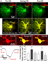
Long lasting spreading depolarization-induced astroglial swelling in the ischemic penumbra-like area at risk following focal photothrombotic stroke. A, MIP images of an astrocyte (asterisk) along with nearby dendrites (arrows) and blood vessels (red; labeled with Texas Red Dextran) in layer I of mouse somatosensory cortex. Each numbered image corresponds with a time point indicated on the respective DC potential recording at the imaging site (top right). Negative deflections in the DC potential represent spreading depolarizations. Note that spontaneous SD is superimposed on a shallow ultraslow DC negativity (asterisk) assumed to be an electrophysiological index of cell death. The dendrites bead during initial photothrombotically-induced spreading depolarization and rapidly recover (<2 min). By contrast, the astrocyte swells with no recovery seen. A spontaneous spreading depolarization occurring 4 min later is accompanied by another round of dendritic beading and recovery (shown at 9 min), while the astrocyte continues to swell. B, Paired single section 2PLSM images showing an astrocyte in the penumbra-like area at risk before (control, red) and after initial photothrombotically-induced spreading depolarization (green). Astroglial process swelling is especially evident. Control and after induced spreading depolarization (Post-SD) images are overlaid (bottom) with arrows pointing to green areas illustrating swelling beyond control morphology. C, Summary from 64 astrocytes in 20 animals showing the increase in astroglial soma size immediately after initial photothrombotically-induced spreading depolarization (post spreading depolarization images were taken 2.3±0.2 min after initial depolarization onset). *p<0.001, paired t-test. D, Somata of EGFP-expressing astrocytes (green dots) and astrocytes labeled with SR101 (red dots) continued to increase in size after initial spreading depolarization onset. Values are shown as percent of control cross-section soma area for each astrocyte. Regression line is shown (black) (r = 0.43, p<0.001; 66 astrocytes from 21 animals). E, Same data as in (D) presented as a bar graph showing a long lasting increase in astroglial soma size following the initial induced spreading depolarization. Values are shown as percent of control for each astrocyte. *p<0.001 relative to control, p<0.05 relative to 0–5, one-way RM ANOVA.
Figure 2
Figure 2
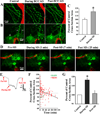
Astroglial swelling in the ischemic penumbra-like area at risk is exacerbated by spontaneous recurrent spreading depolarizations. A, Data from 39 spreading depolarizations in 15 mice showing a positive strong correlation between astroglial soma size and the total duration of spreading depolarization. Values are shown as percent of control, representing the cross-section soma area recorded at 25.1±0.7 min after the onset of the initial spreading depolarization. Regression line is shown (r = 0.64, p<0.001, 37 astrocytes). B, Astroglial soma size increases with occurrence of spontaneous recurrent spreading depolarization. Numbered MIP image sequence of swollen astrocyte (asterisk), dendrites (arrow) and blood vessels (red) taken shortly before (Pre-SD, 1), during (2) and after (Post-SD, 3) spontaneous recurrent spreading depolarization at time points indicated by a corresponding number on the DC potential recording of spontaneous recurrent spreading depolarization shown to the right. Image 2 demonstrates dendritic beading and further astroglial swelling during passage of spontaneous spreading depolarization. Dendrites rapidly recover after spreading depolarization, but astrocyte remains swollen (image 3). Overlays showing the merged Pre-SD/During SD and Pre-SD/Post-SD images reveal green areas (arrows) of additional recurrent spreading depolarization-induced astroglial swelling. C, Summary from 15 astrocytes in 6 animals showing step-wise increase in astroglial soma size during the occurrence of a recurrent spontaneous spreading depolarization. Data points were only used when images were taken immediately prior to and after a recorded recurrent spontaneous spreading depolarization (Pre-SD images were taken 5.3 ± 1.3 min and Post-SD images were taken 2.0 ± 0.3 min after recurrent spreading depolarization onset). *p<0.005, paired t-test.
Figure 3
Figure 3
Astrocytes reversibly swell in response to spreading depolarization in normoxic healthy neocortex. A, Top: MIP images of an astrocyte before, during, and after KCl-induced spreading depolarization (shown in C) in normoxic somatosensory cortex. The astroglial soma and processes swell transiently during spreading depolarization before returning to control morphology. Bottom: Left, overlay showing the merged pre-SD/during SD images for the same astrocyte as above. Center and Right, overlay images of another astrocyte for merged Pre-SD/During SD and for Pre-SD/Post-SD images. Green areas (arrows) represent swelling beyond the pre-SD morphology. Chevron (left image) points to a swollen astroglial endfoot during SD. B, An astrocyte labeled with SR101. Yellow mask showing the Pre-SD cross-section soma area is placed over the same cell During and Post-SD. SD evokes transient swelling (arrows). C, Example spreading depolarization recording from microelectrode placed next to an imaged astrocyte. D, Summary from 15 astrocytes from 5 animals showing reversible astroglial soma swelling in response to two subsequent KCl-induced spreading depolarizations in normoxic somatosensory cortex. Average duration of the first depolarizations was 0.99 ± 0.38 min and the second was 1.73 ± 0.82 min, as measured between the points of half the DC amplitude during depolarization onset and recovery. Astrocytes transiently swell in response to both spreading depolarizations. **p<0.001 and *p<0.02 relative to Pre-SD and Post-SD for each spreading depolarization, one-way RM ANOVA.
Figure 4
Figure 4
Astrocytes swell in response to BCCAO-induced spreading depolarization. A, In vivo 2PLSM high-magnification MIP images of dendrites (green) in layer I of mouse somatosensory cortex along with nearby blood vessel (red). The vessel constricts and dendrites become beaded during BCCAO, coinciding with the occurrence of spreading depolarization. When reperfusion is initiated immediately after the detection of spreading depolarization, dendritic beading is reversed as blood flow returns. B, MIP images of an astrocyte (asterisk) along with nearby dendrites (arrows) and blood vessel (red) in layer I of mouse somatosensory cortex. As in A, the dendrites bead and recover in concert with BCCAO-induced spreading depolarization. The astroglial soma profile increases following BCCAO as its process tree becomes more diffuse. C, Summary from 73 astrocytes in 20 animals showing the increase in astroglial soma size evoked by BCCAO-induced spreading depolarization (during spreading depolarization images were taken 2.3 ± 0.3 min after depolarization onset). *p < 0.001, Wilcoxon Signed Rank Test. D, Images of two astrocytes corresponding to the various time points shown in E during short-lasting spreading depolarization recorded from microelectrode placed next to imaged astrocytes. The astroglial soma and processes swell during short-lasting spreading depolarization and then return to control morphology during post-spreading depolarization reperfusion and DC recovery. F, Data from 9 BCCAO-induced short-lasting spreading depolarizations (4.1±1.0 min average duration) in 9 animals showing that astroglial soma size decreases after an initial swelling. Regression line is shown (black) (r = −0.35, p<0.001; 30 astrocytes). G, Same data as in (F) presented as a bar graph reveal transient astroglial swelling during short-lasting BCCAO-induced spreading depolarization. Images were acquired immediately after onset of spreading depolarization at 2.4±0.6 min (During SD) and after repolarization at 1.7±0.3 min (Post-SD). Data points acquired after repolarization were at 8.8±1.0 min after spreading depolarization onset. Values are shown as percent of control for each astrocyte. *p<0.001, relative to Control and Post-SD, one-way RM ANOVA.
Figure 5
Figure 5
A, Single section image sequence of astrocyte corresponding to the various time points shown on the electrophysiological recording of long-lasting spreading depolarization (left). The astroglial soma and processes swell during spreading depolarization and remain swollen during the late ultraslow potential (asterisk). Continued swelling is also seen at 3 h post-spreading depolarization. B, Data from 13 BCCAO-induced long-lasting spreading depolarizations in 13 animals show that astroglial somata remain swollen after spreading depolarization onset. Values are shown as percent of control, representing the cross-section soma area of each astrocyte. Regression line is shown (black) (r = 0.25, p<0.001, 49 astrocytes). C, Same data as in (B) plotted as a bar graph showing a persistent increase in astroglial soma size following the long-lasting BBCAO-induced spreading depolarization. Values are shown as percent of control cross-section soma area of each astrocyte. *p<0.001 relative to control, one-way RM ANOVA.

Similar articles

Cited by

References

    1. Abdullaev IF, Rudkouskaya A, Schools GP, Kimelberg HK, Mongin AA. Pharmacological comparison of swelling-activated excitatory amino acid release and Cl-currents in cultured rat astrocytes. J Physiol. 2006;572(Pt 3):677–689. - PMC - PubMed
    1. Aitken PG, Tombaugh GC, Turner DA, Somjen GG. Similar propagation of SD and hypoxic SD-like depolarization in rat hippocampus recorded optically and electrically. J Neurophysiol. 1998;80:1514–1521. - PubMed
    1. Amiry-Moghaddam M, Ottersen OP. The molecular basis of water transport in the brain. Nat Rev Neurosci. 2003;4:991–1001. - PubMed
    1. Andrew RD, Labron MW, Boehnke SE, Carnduff L, Kirov SA. Physiological evidence that pyramidal neurons lack functional water channels. Cereb Cortex. 2007;17:787–802. - PubMed
    1. Araque A, Parpura V, Sanzgiri RP, Haydon PG. Tripartite synapses: glia, the unacknowledged partner. Trends Neurosci. 1999;22:208–215. - PubMed

Publication types