. 2012:910:309-35.
doi: 10.1007/978-1-61779-965-5_14.

Models of excitation-contraction coupling in cardiac ventricular myocytes

Affiliations

Models of excitation-contraction coupling in cardiac ventricular myocytes

M Saleet Jafri. Methods Mol Biol. 2012.

Abstract

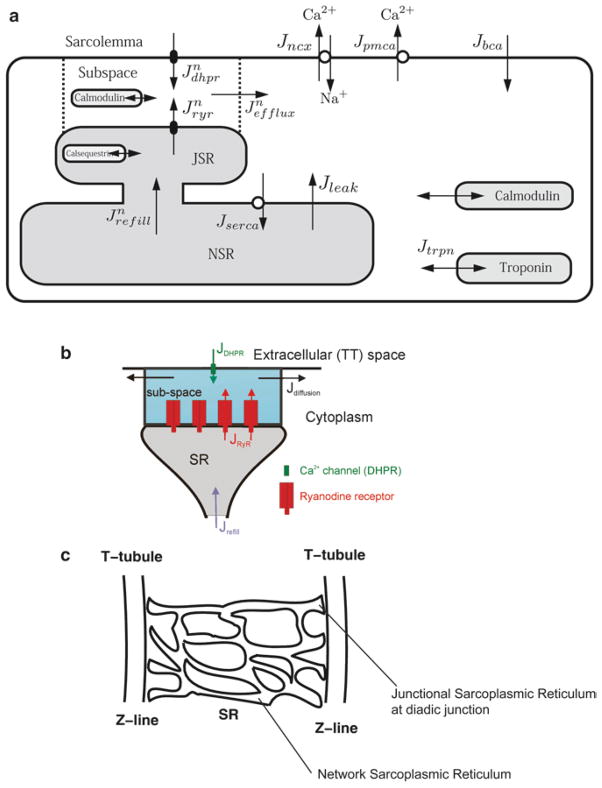
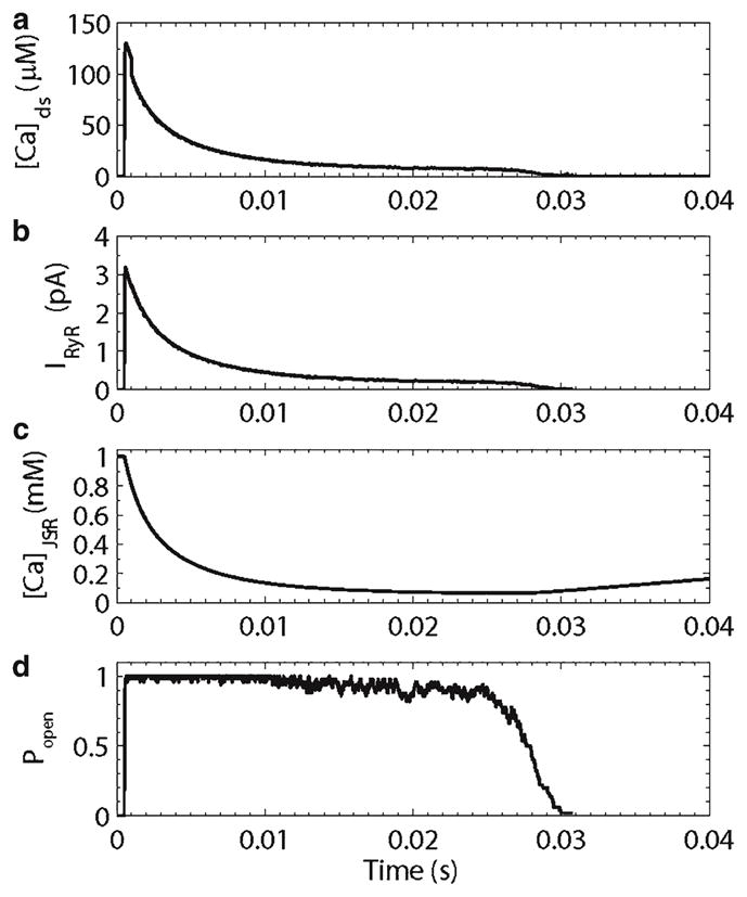
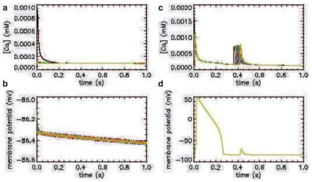
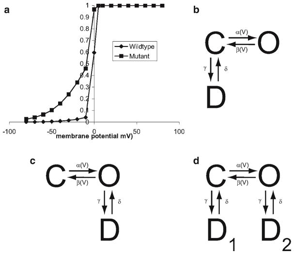
Excitation-contraction coupling describes the processes relating to electrical excitation through force generation and contraction in the heart. It occurs at multiple levels from the whole heart, to single myocytes and down to the sarcomere. A central process that links electrical excitation to contraction is calcium mobilization. Computational models that are well grounded in experimental data have been an effective tool to understand the complex dynamics of the processes involved in excitation-contraction coupling. Presented here is a summary of some computational models that have added to the understanding of the cellular and subcellular mechanisms that control ventricular myocyte calcium dynamics. Models of cardiac ventricular myocytes that have given insight into termination of calcium release and interval-force relations are discussed in this manuscript. Computational modeling of calcium sparks, the elementary events in cardiac excitation-contraction coupling, has given insight into mechanism governing their dynamics and termination as well as their role in excitation-contraction coupling and is described herein.


Figures

Fig. 1
Fig. 1
Schematic diagram of excitation–contraction coupling in the ventricular myocyte. (a) Excitation–contraction coupling model of the guinea pig ventricular myocyte. (b) Model of the calcium release unit in the diad, (c) sarcoplasmic reticulum arrangement in the sarcomere.
Fig. 2
Fig. 2
(a) Circuit diagram of membrane. (b) Simple gating model.
Fig. 3
Fig. 3
(a) The deterministic common pool model of the guinea pig ventricular myocyte does not properly produce graded calcium release. (b) A local control model based on independent stochastic calcium release units that produce calcium sparks successfully produces graded release.
Fig. 4
Fig. 4
(a) State diagram with possible transitions from state Y to state X or Z. (b) The transition probabilities are mapped to the interval [0,1] for comparison to a uniform [0,1] random number.
Fig. 5
Fig. 5
L-type calcium channel model.
Fig. 6
Fig. 6
Schematic diagrams of ryanodine receptor models. (a) Keizer Levine, (b) Smith-Keizer, (c) Sobie et al., (d) the open rate shifts as the sarcoplasmic reticulum depletes in the Sobie et al. model.
Fig. 7
Fig. 7
Simulated excitation–contraction coupling in the guinea pig ventricular myocyte. (a) Action potential, (b) myoplasmic calcium transient, (c) isometric force transient.
Fig. 8
Fig. 8
Interval–force relations. (a) Peak myoplasmic calcium, (b) peak sarcoplasmic reticulum calcium concentration, (c) peak ryanodine receptor open probability, (d) peak ryanodine receptor release flux.
Fig. 9
Fig. 9
Simulated calcium spark. (a) Diadic subspace calcium concentration, (b) ryanodine receptor release current, (c) junctional sarcoplasmic reticulum calcium concentration, (d) ryanodine receptor open probability.
Fig. 10
Fig. 10
Simulated calcium and membrane potential transients (top and bottom rows, respectively) for a one-dimensional spatial model of a normal and pathological guinea pig ventricular myocyte (left and right columns, respectively).
Fig. 11
Fig. 11
(a) Wild-type and mutant steady-state activation curves for a hypothetical channel. (b) Nonuse-dependent block by drug. (c) Use-dependent block by drug. (d) Use-independent block by drug.

References

    1. Hodgkin AL, Huxley AF. A quantitative description of membrane current and its application to conduction and excitation in nerve. J Physiol. 1952;117:500–544. - PMC - PubMed
    1. Williams GS, Smith GD, Sobie EA, Jafri MS. Models of cardiac excitation–contraction coupling in ventricular myocytes. Math Biosci. 2010;226:1–15. - PMC - PubMed
    1. DiFrancesco D, Noble D. A model of cardiac electrical activity incorporating ionic pumps and concentration changes. Philos Trans R Soc Lond B Biol Sci. 1985;307:353–398. - PubMed
    1. Beeler GW, Reuter H. Reconstruction of the action potential of ventricular myocarial fibres. J Physiol. 1977;268:177–210. - PMC - PubMed
    1. Luo CH, Rudy Y. A model of the ventricular cardiac action potential. Depolarization, repolarization, and their interaction. Circ Res. 1991;68:1501–1526. - PubMed

MeSH terms

LinkOut - more resources