. 2013 Jan;57(1):228-38.
doi: 10.1002/hep.25983.

Scavenger receptor A restrains T-cell activation and protects against concanavalin A-induced hepatic injury

Affiliations

Scavenger receptor A restrains T-cell activation and protects against concanavalin A-induced hepatic injury

Daming Zuo et al. Hepatology. 2013 Jan.

Abstract

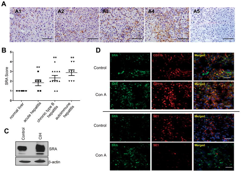
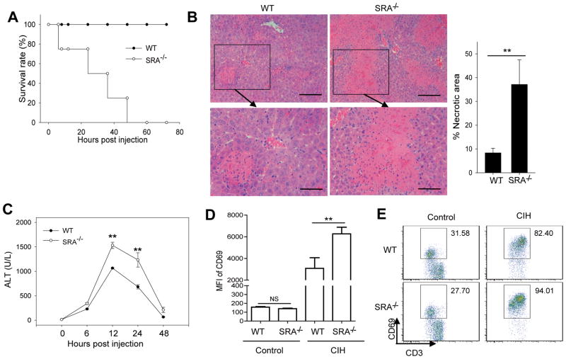
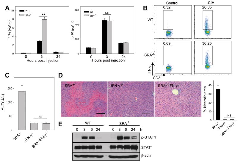
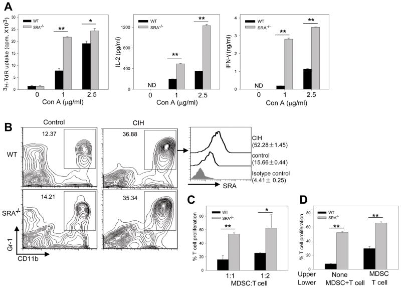
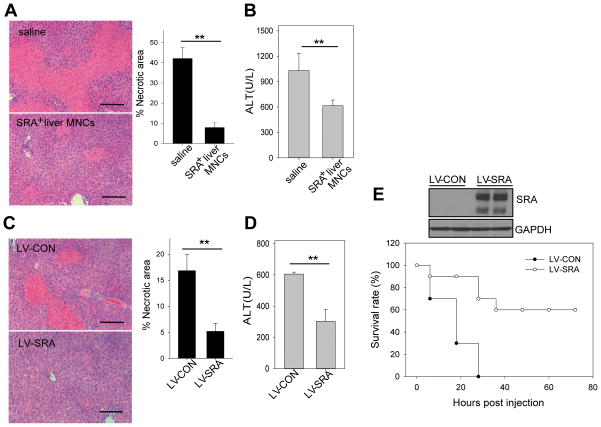
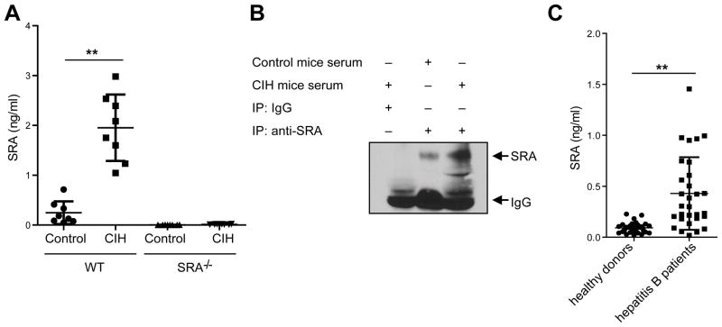
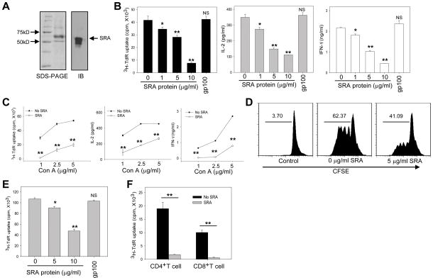
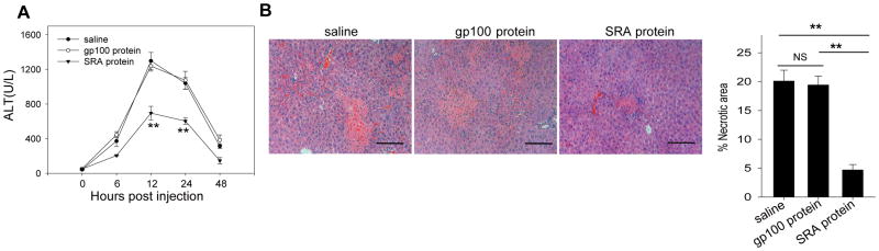
Negative feedback immune mechanisms are essential for maintenance of hepatic homeostasis and prevention of immune-mediated liver injury. We show here that scavenger receptor A (SRA/CD204), a pattern recognition molecule, is highly up-regulated in the livers of patients with autoimmune or viral hepatitis, and of mice during concanavalin A (Con A)-induced hepatitis (CIH). Strikingly, genetic SRA ablation strongly sensitizes mice to Con A-induced liver injury. SRA loss, increased mortality and liver pathology correlate with excessive production of IFN-γ and heightened activation of T cells. Increased liver expression of SRA primarily occurs in mobilized hepatic myeloid cells during CIH, including CD11b(+) Gr-1(+) cells. Mechanistic studies establish that SRA on these cells functions as a negative regulator limiting T-cell activity and cytokine production. SRA-mediated protection from CIH is further validated by adoptive transfer of SRA(+) hepatic mononuclear cells or administration of a lentivirus-expressing SRA, which effectively ameliorates Con A-induced hepatic injury. Also, CIH and clinical hepatitis are associated with increased levels of soluble SRA. This soluble SRA displays a direct T-cell inhibitory effect and is capable of mitigating Con A-induced liver pathology.

Conclusion: Our findings demonstrate an unexpected role of SRA in attenuation of Con A-induced, T-cell-mediated hepatic injury. We propose that SRA serves as an important negative feedback mechanism in liver immune homeostasis, and may be exploited for therapeutic treatment of inflammatory liver diseases.


Figures

Figure 1
Figure 1. Upregulation of SRA in human hepatitis and the mouse model of experimental hepatitis
(A) Representative immunohistochemical staining of liver sections for SRA expression. (A1) normal human liver; (A2) acute viral hepatitis; (A3) chronic type B hepatitis; (A4) autoimmune hepatitis; (A5) Normal serum staining from A4. (B) Slides were examined and scored independently by the pathologists in a blinded fashion. SRA expression was quantitated using a grading system based on the intensity of staining and classified into four groups: 0 (undetected), 1 (very low), 2 (low), 3 (high), 4 (very high). p<0.01, compared to normal human liver. (C) Immunoblotting analysis of hepatic SRA expression in WT mice with or without Con A (15 mg/kg) treatment. (D). Expression pattern of SRA on myeloid cells and LSECs during CIH. Liver sections from control mice or those receiving Con A (n=5) were subjected to double staining with anti-SRA and anti-CD11b or anti-SE1 Abs. DAPI was used to counterstain nucleus (scale bar, 50 μm).
Figure 2
Figure 2. SRA ablation renders mice more susceptible to CIH
(A) WT and SRA−/−mice (n=8) were injected with Con A at the dose of 25 mg/kg, and overall survival of mice was determined. (B) 20 hours after Con A injection (15 mg/kg), histological analysis of mouse livers (n=5) was performed using H&E staining. Bottom panels show the higher magnification views of the necrotic area. Scale bar (top), 100 μm; Scare bar (bottom), 50 μm. The percentage of necrotic area was quantitated using ImageJ software. (C) Serum ALT levels were assessed at the different time points as indicated following Con A (15 mg/kg) injection. (D, E) SRA ablation results in increased T cell activation. Mean fluorescence intensity (MFI) of CD69 on T cells and the percentage of CD69+ T cells before and after Con A injection is assessed by FACS analysis of liver MNCs. ** p<0.01; NS, not significant. Data are representative of three independent experiments with similar results.
Figure 3
Figure 3. SRA ablation leads to increased IFN-γ production during CIH
(A) Serum levels of IFN-γ and IL-10 were determined at 3 and 24 hours after Con A (15 mg/kg) injection to mice (n=5). (B) Increased frequency of IFN-γ-producing T cells, analyzed 14 hours after Con A injection using intracellular cytokine staining. (C, D) IFN-γ deficiency abolishes SRA loss-promoted CIH. SRA−/−, IFN-γ−/−and SRA−/−IFN-γ−/−mice (n=3) were injected with Con A. ALT levels (C) and liver histology (D) were examined 20 hours after Con A injection (scale bar, 100 μm). Necrotic area was quantitated and calculated based on at least 30 random fields from the liver samples of three mice. (E) Increased STAT-1 activation in the livers from Con A-treated SRA−/−mice. Levels of phospho-STAT1 and total STAT1 protein were analyzed using immunoblotting. ** p<0.01; NS, not significant. The data shown represent two independent experiments.
Figure 4
Figure 4. Upregulation of SRA on hepatic CD11b+Gr-1+ MDSCs correlates with their immunosuppressive activity during CIH
(A) SRA−/−hepatic MNCs are more responsive to Con A stimulation. Hepatic MNCs (4 × 10⁵ per well) were cultured with Con A at different concentrations. T cell proliferation and cytokine levels were determined by 3H-thymidine incorporation and ELISA assays, respectively. Data are representative of three independent experiments. ** p<0.01; * p<0.05; ND, not detectable. (B) Elevation of SRA expression on hepatic CD11b+Gr-1+ myeloid cells. Hepatic MNCs from WT and SRA−/−mice (n=5) were stained for CD11b and Gr-1, followed by FACS analysis (left). SRA expression on CD11b+Gr-1+ cells from WT mice, as indicated by MFI, was also examined (right). (C) SRA absence on hepatic MDSCs reduces their immunosuppressive activity. Splenocytes were stimulated with anti-CD3/CD28 Abs in the presence of hepatic MDSCs isolated from Con A-injected WT and SRA−/−mice. (D) SRA-mediated immune suppression by MDSCs requires interaction with T cells. T cells were co-cultured with MDSCs in the lower chambers of the transwell plates, or separated from MDSCs by seeding MDSCs to the upper chambers. T cell proliferation in the absence of MDSC in (C and D) was set as 100 %. * p<0.05; ** p<0.01. All samples were run in triplicate. Data shown represent two independent experiments.
Figure 5
Figure 5. Increased levels of hepatic SRA expression protect mice from CIH
(A, B) Adoptive transfer of SRA+ hepatic MNCs ameliorates CIH in SRA−/−mice. SRA−/−mice (n=5) were injected with WT hepatic MNCs (5×10⁶ cells) 2 hours before Con A (15 mg/kg) injection. Mice were examined 20 hours later for liver histology (A) and serum ALT levels (B). (C, D) Lentiviral-mediated overexpression of SRA reduces CIH in WT mice. WT mice (n=5) were i.v. injected with 2×10⁷ TU LV-CON or LV-SRA 6 days and 3 days before Con A (15 mg/kg) administration. Liver histology (C) and ALT levels (D) were examined. Scale bar, 100 μm. (E) Enhanced survival of Con A-challenged SRA−/−mice by upregulation of hepatic SRA. SRA−/−mice (n=10) were injected with LV-SRA or LV-CON before Con A (25 mg/kg) administration. Lentivirus-mediated hepatic SRA expression was examined prior to Con A injection by immunobloting. ** p<0.01. Data shown are representative of three independent experiments.
Figure 6
Figure 6. Liver injury is associated with soluble SRA
(A) WT mice (n=8) were injected with Con A (15 mg/kg). Serum levels of SRA were measured 24 hours later by ELISA. Sera collected from SRA−/−mice before and after Con A injection were used as negative controls. (B) The presence of SRA in sera was confirmed by immunoprecipitation assays using anti-SRA Abs. ** p<0.01. Data represent three independent experiments. (C) Presence of soluble SRA in patients with hepatitis B virus (n=30) compared to healthy donors (n=30).
Figure 7
Figure 7. Extracellular SRA protein inhibits T cell activation
(A) Preparation of recombinant SRA protein using baculovirus-insect cell system, analyzed by SDS-PAGE and immunoblotting. (B) Reduced hepatic MNC proliferation by SRA protein. 4×10⁵ purified MNCs were stimulated with Con A (2.5 μg/ml) in the presence of indicated concentrations of SRA protein. Cell proliferation and cytokine production were determined. gp100 protein was used as an irrelevant protein control. (C) Effect of extracellular SRA (10 μg/ml) on hepatic MNCs proliferation-simulated by Con A at different concentrations. (D) Inhibition of Con A-induced T cell proliferation by SRA protein. CFSE (5 μM)-labeled hepatic MNCs were stimulated with Con A in the presence of SRA protein for 72 hours. T cell proliferation was assessed by FACS analysis of CD3+ T cells based on the dilution of CFSE intensity. Representative histograms from two independent experiments are shown. (E) Purified T cells were stimulated with anti-CD3/CD28 Abs in the presence of SRA protein, followed by 3H-thymidine incorporation assays. (F) Proliferation of purified CD4+ or CD8+ T cells in the presence of SRA protein. * p<0.05; ** p<0.01; NS, not significant, compared to the group without SRA protein. All samples were run in triplicate. Data shown represent three independent experiments.
Figure 8
Figure 8. Soluble SRA protein partially protects mice from CIH
WT mice (n=5) received two doses of SRA protein (200 μg) 1 hour before and 2 hours after Con A (15 mg/kg) injection. gp100 protein was used as an irrelevant protein control. Serum ALT (A) and histology of liver (B) were examined 20 hours following Con A administration. Quantitative data of the percentage of necrotic area are also presented. Scale bar, 100 μm. ** p<0.01. Data are representative of at least three independent experiments.

References

    1. Dienes HP, Drebber U. Pathology of immune-mediated liver injury. Dig Dis. 2010;28:57–62. - PubMed
    1. Lohr HF, Schlaak JF, Gerken G, Fleischer B, Dienes HP, Meyer zum Buschenfelde KH. Phenotypical analysis and cytokine release of liver-infiltrating and peripheral blood T lymphocytes from patients with chronic hepatitis of different etiology. Liver. 1994;14:161–166. - PubMed
    1. McFarlane IG. Pathogenesis of autoimmune hepatitis. Biomed Pharmacother. 1999;53:255–263. - PubMed
    1. Rehermann B. Intrahepatic T cells in hepatitis B: viral control versus liver cell injury. J Exp Med. 2000;191:1263–1268. - PMC - PubMed
    1. Tiegs G, Hentschel J, Wendel A. A T cell-dependent experimental liver injury in mice inducible by concanavalin A. J Clin Invest. 1992;90:196–203. - PMC - PubMed

Publication types