

Science to practice: how will myocardial inflammation be imaged with MR imaging?
- PMID: 22821689
- PMCID: PMC6939999
- DOI: 10.1148/radiol.12121094
Science to practice: how will myocardial inflammation be imaged with MR imaging?
Abstract
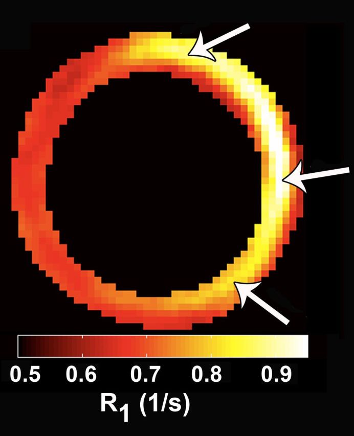
The elegant study by Naresh and colleagues (1) synthesizes many of the best aspects of molecular magnetic resonance(MR) imaging: Quantitative serial imaging of a well-defined molecular process is performed in vivo, and its results are correlated with sensitive measures of left ventricular function. The technique described adds a valuable tool to the molecular imaging armamentarium. How, then, will myocardial inflammation be imaged with MR imaging?The only clinical experience to date has been with iron oxide nanoparticles (2,3). Their excellent sensitivity, dynamic range, and safety record make them a highly appealing choice. It will be critical, however, for any iron oxide nanoparticle that is used clinically to be well studied and validated in animal models of the disease before it is used in humans. A “group effect” cannot be assumed, even in the case of fairly similar iron oxide nanoparticles. The use of MR imaging–detectable liposomes appears promising,and initial clinical studies with fluorine-containing liposomes are likely to begin shortly. The clinical use of gadolinium-labeled liposomes appears further away, and the approach described by Naresh and colleagues is thus likely to remain confined to preclinical investigation for the foreseeable future. The development of novel anti-inflammatory therapies, however, will require robust imaging tools to shepherd these agents through preclinical studies and into the clinical arena. The approach described by Naresh et al adds a valuable tool to the preclinical molecular imaging armamentarium.
Comment on
-
Monocyte and/or macrophage infiltration of heart after myocardial infarction: MR imaging by using T1-shortening liposomes.Radiology. 2012 Aug;264(2):428-35. doi: 10.1148/radiol.12111863. Epub 2012 Jun 21. Radiology. 2012. PMID: 22723500 Free PMC article.
References
-
- Harisinghani MG, Barentsz J, Hahn PF, et al. Noninvasive detection of clinically occult lymph-node metastases in prostate cancer. N Engl J Med 2003;348(25):2491–2499. - PubMed
-
- Tang TY, Howarth SP, Miller SR, et al. The ATHEROMA (Atorvastatin Therapy: Effects on Reduction of Macrophage Activity) Study: evaluation using ultrasmall superparamagnetic iron oxide-enhanced magnetic resonance imaging in carotid disease. J Am Coll Cardiol 2009;53(22):2039–2050. - PubMed
-
- Sosnovik DE, Nahrendorf M, Deliolanis N, et al. Fluorescence tomography and magnetic resonance imaging of myocardial macrophage infiltration in infarcted myocardium in vivo. Circulation 2007;115(11):1384–1391. - PubMed
Publication types
MeSH terms
Substances
Grants and funding
LinkOut - more resources
Full Text Sources
Medical
